
CBCT Imaging at AS Medical Complex
Contact hospital
- Availability
Contact hospital
- Booking
WhatsApp / call
- Location
Islamabad
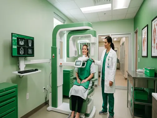
Cone Beam Computed Tomography for Advanced Dental & Maxillofacial Imaging
The CBCT Service at AS Medical Complex provides three-dimensional diagnostic imaging for selected dental, oral, maxillofacial, orthodontic and related conditions where conventional two-dimensional X-rays may not provide sufficient information.
CBCT stands for Cone Beam Computed Tomography.
It is a specialized X-ray-based imaging technique in which a cone-shaped X-ray beam rotates around the patient and collects information that is reconstructed by computer into a three-dimensional image.
CBCT may provide detailed visualization of:
-
Teeth
-
Tooth roots
-
Jaw bones
-
Alveolar bone
-
Impacted teeth
-
Dental implant sites
-
Root-canal anatomy
-
Temporomandibular joints
-
Maxillary sinuses
-
Nasal cavity
-
Selected facial bones
-
Nerve canals
-
Other dentofacial structures
The FDA identifies dental implant planning, evaluation of abnormal or impacted teeth, jaw and facial assessment, cleft-palate assessment, selected caries evaluation, endodontic diagnosis and dental-trauma assessment among established clinical applications of dental CBCT.
At AS Medical Complex, CBCT should be requested when its three-dimensional information is expected to improve:
-
Diagnosis
-
Treatment planning
-
Surgical planning
-
Anatomical assessment
-
Evaluation of complex disease
-
Follow-up in selected cases
CBCT should not automatically replace routine dental X-rays.
FDA guidance recommends performing dental CBCT only when the necessary clinical information cannot be adequately obtained using another imaging method with less or no ionizing radiation.
Our CBCT Services
Depending on clinical indication, scanner capability and available reporting expertise, CBCT services at AS Medical Complex may include:
-
Dental CBCT
-
Maxillofacial CBCT
-
CBCT of the upper jaw
-
CBCT of the lower jaw
-
Full-jaw CBCT where clinically appropriate
-
Limited Field of View CBCT
-
Dental implant planning
-
Pre-implant bone assessment
-
Assessment of implant sites
-
Measurement of available jaw bone
-
Identification of relevant anatomical structures
-
Inferior alveolar nerve-canal assessment
-
Mental foramen assessment
-
Maxillary-sinus assessment before dental treatment
-
Impacted wisdom-tooth assessment
-
Impacted canine assessment
-
Other impacted-tooth assessment
-
Tooth-root morphology assessment
-
Endodontic CBCT in selected complex cases
-
Root-canal anatomy assessment
-
Assessment for selected missed canals
-
Periapical-lesion assessment
-
Selected root-resorption assessment
-
Selected root-fracture assessment
-
Dental-trauma imaging
-
Jaw-lesion assessment
-
Selected cyst assessment
-
Selected jaw-tumor assessment
-
Temporomandibular-joint bony assessment
-
Orthodontic assessment where clinically indicated
-
Cephalometric-related 3D assessment where available
-
Selected facial-bone assessment
-
Cleft and craniofacial assessment where appropriate
-
Selected nasal-cavity assessment
-
Selected paranasal-sinus assessment
-
Preoperative oral-surgery planning
-
Selected postoperative assessment
-
Referral for oral and maxillofacial radiology interpretation where necessary
Specialized CBCT applications should only be advertised as routine onsite services after confirming the exact scanner, field-of-view options, software and reporting expertise available at AS Medical Complex.
What Is CBCT?
Cone Beam Computed Tomography is a form of computed tomography designed particularly for high-resolution imaging of structures within the dental and maxillofacial region.
During the examination:
-
The patient is positioned in the CBCT unit.
-
The X-ray source and detector rotate around the head.
-
A cone-shaped X-ray beam collects imaging data.
-
Computer software reconstructs the information into a three-dimensional volume.
The resulting dataset can be viewed in multiple planes.
These may include:
-
Axial
-
Coronal
-
Sagittal
-
Oblique
-
Curved panoramic
-
Three-dimensional reconstructed views
FDA describes CBCT as a variation of conventional CT in which a cone-shaped X-ray beam is used to reconstruct three-dimensional images of dental, oral, maxillofacial and selected ENT anatomy.
CBCT vs Conventional Dental X-Ray
Routine dental X-rays usually provide two-dimensional information.
Examples include:
-
Periapical X-Ray
-
Bitewing X-Ray
-
Panoramic radiography
-
Other conventional dental projections
These examinations remain appropriate for many routine dental conditions.
CBCT provides three-dimensional information.
This can reduce the problem of anatomical structures overlapping one another on a conventional two-dimensional image.
FDA notes that one of CBCT’s main advantages is the ability to provide 3D rather than 2D anatomical information.
However, CBCT generally exposes the patient to more radiation than conventional dental radiography.
It should therefore be used when the additional information is expected to influence diagnosis or treatment.
CBCT vs Conventional CT Scan
CBCT and conventional medical CT both create cross-sectional imaging, but they are not identical examinations.
CBCT
CBCT is particularly suited to high-resolution assessment of:
-
Teeth
-
Jaw bones
-
Dental implant sites
-
Tooth roots
-
Bony maxillofacial anatomy
-
Selected sinuses
-
Selected TMJ structures
Conventional CT
Medical CT may provide better assessment of:
-
Extensive facial trauma
-
Deep neck disease
-
Soft tissues
-
Tumor spread
-
Lymph nodes
-
Major infections
-
Other complex head and neck conditions
RadiologyInfo notes that CBCT provides excellent detail of bone and dental structures but does not provide the same soft-tissue diagnostic information as conventional CT for structures such as muscles, glands and lymph nodes.
Therefore, CBCT should not simply replace conventional CT whenever head or neck imaging is required.
Three-Dimensional Dental Imaging
A major benefit of CBCT is its ability to demonstrate the relationship between structures in three dimensions.
For example, a dentist or oral surgeon may need to understand the relationship between:
-
A wisdom tooth and the mandibular nerve canal
-
An implant site and the maxillary sinus
-
A tooth root and surrounding bone
-
An impacted canine and adjacent tooth roots
-
A jaw lesion and surrounding structures
This information may not always be fully appreciated on a two-dimensional dental X-ray.
Field of View
The Field of View – FOV refers to the amount of anatomy included within the CBCT scan.
A CBCT examination may use:
-
Small Field of View
-
Medium Field of View
-
Larger Field of View
depending on the clinical question and scanner.
A small field may be appropriate when only one tooth or a small region needs detailed assessment.
A larger field may be required for:
-
Multiple implant sites
-
Orthodontic planning
-
Larger jaw lesions
-
Complex maxillofacial assessment
Radiation exposure should be optimized by limiting the scanned area to the anatomy required for diagnosis whenever possible. FDA specifically recommends selecting exposure settings according to clinical indication, patient size and anatomical region while keeping radiation as low as reasonably achievable.
CBCT for Dental Implant Planning
Dental implant placement is one of the major applications of CBCT.
CBCT may help evaluate:
-
Jaw-bone height
-
Jaw-bone width
-
Implant-site anatomy
-
Relationship to adjacent teeth
-
Inferior alveolar nerve canal
-
Mental foramen
-
Maxillary sinus
-
Nasal cavity
-
Other nearby structures
FDA identifies dental implant planning as an established CBCT application.
Bone Assessment Before Dental Implants
Successful implant planning requires an understanding of the available supporting bone.
CBCT may allow the clinician to assess:
-
Bone dimensions
-
Ridge shape
-
Anatomical limitations
-
Selected bony defects
-
Relationship with important structures
However, CBCT alone does not determine whether a patient is suitable for an implant.
Clinical assessment should also consider:
-
Gum health
-
Oral hygiene
-
Smoking
-
Diabetes
-
Medication
-
Medical conditions
-
Previous dental infection
-
Other relevant factors
Inferior Alveolar Nerve Canal
The inferior alveolar nerve travels through the lower jaw.
Its location can be important when planning:
-
Dental implants
-
Wisdom-tooth surgery
-
Other mandibular procedures
CBCT may help demonstrate the three-dimensional relationship between the planned surgical area and the nerve canal.
This can support treatment planning and risk assessment.
Mental Foramen
The mental foramen is an opening in the lower jaw through which a nerve and blood vessels emerge.
Its location may be important during implant or oral-surgical planning.
CBCT can help identify its relationship to the planned treatment site.
Maxillary Sinus & Implant Planning
The maxillary sinuses lie above the upper back teeth.
When implants are being considered in the posterior upper jaw, CBCT may help assess:
-
Bone height below the sinus
-
Sinus anatomy
-
Relationship of tooth roots to the sinus
-
Selected sinus abnormalities
-
Need for additional surgical planning
RadiologyInfo identifies evaluation of the jaw and maxillary sinuses as an important CBCT application.
Sinus Lift Planning
Some patients have insufficient bone height in the posterior upper jaw for straightforward implant placement.
A dentist or oral surgeon may consider a sinus augmentation or sinus-lift procedure in selected cases.
CBCT may help with preoperative assessment of:
-
Available bone
-
Sinus anatomy
-
Relevant anatomical structures
The decision to perform sinus augmentation requires specialist dental or oral-surgical assessment.
Impacted Wisdom Teeth
Wisdom teeth may fail to erupt normally and remain partially or completely impacted.
CBCT may be useful in selected complex cases to assess:
-
Tooth orientation
-
Number and shape of roots
-
Relationship to adjacent teeth
-
Relationship to the mandibular nerve canal
-
Surrounding bone
-
Other anatomical structures
FDA includes visualization of abnormal teeth among established CBCT applications.
Not every wisdom tooth requires CBCT.
Many can be adequately evaluated using clinical examination and conventional dental radiography.
Wisdom Tooth & Nerve Relationship
For lower wisdom teeth, one important concern is the relationship between tooth roots and the inferior alveolar nerve canal.
In selected cases where conventional X-rays suggest close proximity, CBCT may provide additional three-dimensional information.
This can help an oral surgeon discuss:
-
Surgical approach
-
Potential nerve-related risk
-
Alternative treatment options
CBCT does not eliminate the possibility of nerve injury but can improve anatomical assessment.
Impacted Canines
Upper canine teeth can sometimes remain impacted.
CBCT may help determine:
-
Exact tooth position
-
Orientation
-
Relationship to nearby roots
-
Available eruption pathway
-
Associated damage such as selected root resorption
This information may assist:
-
Orthodontic planning
-
Surgical exposure planning
-
Combined orthodontic-surgical treatment
RadiologyInfo identifies surgical planning for impacted teeth as a common CBCT use.
Orthodontic CBCT
CBCT may be used in selected orthodontic cases when three-dimensional information is required.
Possible indications may include:
-
Impacted teeth
-
Complex tooth position
-
Craniofacial abnormalities
-
Cleft palate
-
Selected skeletal abnormalities
-
Complex surgical-orthodontic planning
-
Selected airway or skeletal assessments
FDA identifies orthodontic-related assessment and cleft-palate evaluation among clinical CBCT applications.
CBCT should not automatically be performed for every orthodontic patient.
Endodontic CBCT
Endodontics involves diagnosis and treatment of conditions affecting the dental pulp and tissues surrounding tooth roots.
CBCT can provide detailed three-dimensional information in selected complex endodontic cases.
Possible applications may include:
-
Complex root anatomy
-
Suspected missed canals
-
Periapical disease
-
Root resorption
-
Selected root fractures
-
Previous root-canal treatment failure
-
Endodontic surgery planning
-
Other difficult diagnostic situations
The updated AAE/AAOMR guidance emphasizes that CBCT should not be used routinely for endodontic screening and should be selected when the required information cannot be adequately obtained from lower-dose two-dimensional radiographs.
Root Canal Anatomy
Teeth can have complex root and canal anatomy.
CBCT may help identify selected anatomical features such as:
-
Additional roots
-
Additional canals
-
Unusual root curvature
-
Complex internal anatomy
This can be helpful in selected difficult root-canal cases.
However, a CBCT should not automatically be ordered before every routine root canal.
Missed Root Canals
Persistent symptoms after previous root-canal treatment can occasionally result from untreated or missed canal anatomy.
In selected cases, CBCT may help identify anatomy that was difficult to appreciate on conventional X-rays.
The dentist or Endodontist must interpret the imaging together with:
-
Symptoms
-
Clinical examination
-
Previous treatment
-
Conventional radiographs
Periapical Lesions
A periapical lesion is an abnormal area surrounding the tip of a tooth root.
Possible causes include:
-
Dental-pulp infection
-
Previous root-canal disease
-
Other dental conditions
CBCT may provide more detailed information about selected lesions than a two-dimensional radiograph.
However, not every periapical abnormality requires CBCT.
Root Resorption
Root resorption occurs when parts of the tooth root are lost through biological processes.
CBCT may help assess selected cases of:
-
Internal resorption
-
External resorption
-
Relationship to surrounding structures
-
Extent of the defect
Detailed three-dimensional information can support treatment planning in complex cases.
Root Fractures
Some tooth-root fractures can be difficult to diagnose.
CBCT may provide useful information in selected cases, although image artifacts and fracture orientation can still affect visibility.
AAE guidance notes that CBCT has an important role in selected complex endodontic diagnostic situations rather than routine use for every patient.
Dental Trauma
Dental trauma can result from:
-
Falls
-
Sports injuries
-
Road accidents
-
Direct facial injury
-
Other trauma
CBCT may be considered in selected complex cases involving:
-
Tooth displacement
-
Root fracture
-
Alveolar-bone injury
-
Impacted or displaced teeth
-
Other dentoalveolar injury
FDA identifies dental-trauma diagnosis as a recognized CBCT application.
Severe facial trauma may require conventional medical CT rather than dental CBCT.
Jaw Cysts
CBCT may help evaluate selected cystic lesions involving the jaws.
It can provide information about:
-
Size
-
Location
-
Relationship to teeth
-
Relationship to the nerve canal
-
Cortical bone changes
-
Other anatomical relationships
However, imaging appearance alone may not establish the exact diagnosis.
Some lesions require:
-
Biopsy
-
Histopathology
-
Oral surgery
-
Additional CT or MRI
Jaw Tumors
CBCT can demonstrate selected bony jaw tumors or tumor-like abnormalities.
RadiologyInfo identifies detection and measurement of jaw tumors among potential CBCT uses.
A suspicious jaw lesion may require additional evaluation through:
-
Clinical examination
-
Biopsy
-
Pathology
-
Conventional CT
-
MRI
-
Specialist maxillofacial assessment
CBCT should not be considered a substitute for tissue diagnosis when biopsy is required.
CBCT & Oral Cancer
CBCT is not a general screening test for oral cancer.
Many oral cancers begin within soft tissues that may not be adequately characterized by CBCT.
Suspicious symptoms such as:
-
Persistent mouth ulcer
-
Unexplained oral lump
-
Persistent red or white patch
-
Unexplained bleeding
-
Persistent swallowing difficulty
-
Unexplained neck lump
require direct clinical assessment.
Further evaluation may involve:
-
Biopsy
-
Pathology
-
CT
-
MRI
-
ENT or maxillofacial referral
depending on the condition.
Temporomandibular Joint – TMJ
The temporomandibular joints connect the lower jaw to the skull.
CBCT can provide detailed information about the bony components of the TMJ.
Possible applications may include selected assessment of:
-
Degenerative bony changes
-
Condylar abnormalities
-
Fracture
-
Ankylosis
-
Other structural changes
RadiologyInfo includes TMJ assessment among common CBCT applications.
TMJ Pain
Patients with TMJ-related symptoms may experience:
-
Jaw pain
-
Clicking
-
Restricted opening
-
Difficulty chewing
-
Facial discomfort
CBCT is not required for every patient with TMJ pain.
Clinical evaluation usually comes first.
MRI may be preferred when the main concern involves:
-
Articular disc
-
Joint soft tissues
-
Other non-bony structures
CBCT and MRI therefore answer different TMJ questions.
Maxillofacial Imaging
CBCT may provide detailed bony imaging of selected structures within the maxillofacial region.
This can include:
-
Maxilla
-
Mandible
-
Teeth
-
Alveolar bone
-
Selected facial bones
-
Sinuses
-
Nasal cavity
The appropriate imaging method depends on whether the primary concern involves:
-
Bone
-
Teeth
-
Soft tissue
-
Trauma
-
Tumor
-
Infection
Facial Fractures
CBCT may demonstrate selected facial-bone fractures.
However, patients with:
-
Major facial trauma
-
Suspected multiple fractures
-
Significant soft-tissue injury
-
Head injury
-
Major bleeding
may require emergency conventional CT Scan rather than an outpatient dental CBCT examination.
Cleft Palate & Craniofacial Conditions
CBCT can contribute to selected assessment of craniofacial and cleft-related bony anatomy.
FDA lists cleft-palate assessment among recognized applications of dental CBCT.
Complex patients may require multidisciplinary assessment involving:
-
Dentistry
-
Orthodontics
-
Plastic Surgery
-
ENT
-
Oral and maxillofacial specialists
-
Other appropriate services
CBCT & ENT
CBCT can include selected ENT-related bony anatomy such as:
-
Paranasal sinuses
-
Nasal cavity
-
Adjacent facial structures
FDA specifically includes ears, nose and throat anatomy among areas that dental CBCT systems may reconstruct.
However, CBCT should not automatically replace conventional sinus CT or other ENT imaging.
The appropriate examination depends on the clinical question and required soft-tissue detail.
Sinus Assessment
CBCT may demonstrate:
-
Maxillary-sinus anatomy
-
Selected mucosal changes
-
Relationship between dental roots and sinus floor
-
Selected bony sinus abnormalities
This may be useful when investigating:
-
Implant planning
-
Dental disease extending toward the sinus
-
Selected ENT-related concerns
A sinus abnormality on CBCT should be correlated with symptoms and specialist assessment.
Dental Infection & Maxillary Sinus
Upper back tooth roots can lie close to the maxillary sinus.
In selected patients, dental infection may be associated with sinus disease.
CBCT may help demonstrate:
-
Tooth-root anatomy
-
Periapical disease
-
Relationship to sinus floor
-
Adjacent bony changes
Management may require coordination between:
-
Dentistry
-
Endodontics
-
ENT
depending on the condition.
Airway Assessment
Some CBCT systems and software can provide three-dimensional visualization of portions of the upper airway.
However, static CBCT airway measurements should not automatically be treated as a diagnosis of sleep apnea.
Sleep apnea diagnosis usually requires appropriate:
-
Clinical assessment
-
Sleep evaluation
-
Sleep study where indicated
Airway analysis should therefore be used cautiously and only for appropriate clinical purposes.
Periodontal Bone Assessment
Periodontal disease can lead to loss of bone supporting the teeth.
CBCT may provide three-dimensional information about selected complex bony defects.
However, routine periodontal diagnosis often relies on:
-
Clinical periodontal examination
-
Probing
-
Conventional dental radiographs
CBCT should be reserved for situations where additional 3D information is expected to change treatment planning.
Bone Grafting
Patients undergoing selected dental bone-grafting procedures may require CBCT before or after treatment.
CBCT may help assess:
-
Ridge anatomy
-
Bone volume
-
Relevant structures
-
Surgical site
The exact imaging schedule should be determined by the treating dental surgeon.
Guided Implant Surgery
CBCT data may be incorporated into digital implant-planning workflows.
Depending on available systems, it can help clinicians plan:
-
Implant position
-
Implant angle
-
Implant depth
-
Relationship to critical anatomy
Some practices combine CBCT data with digital dental scans to produce surgical guides.
This should only be advertised at AS Medical Complex if the required digital planning and surgical-guidance services are actually available.
CBCT Before Extraction
CBCT is not required before every tooth extraction.
It may be considered when:
-
Anatomy is complex
-
A tooth is deeply impacted
-
Important anatomical structures are nearby
-
Conventional radiographs are insufficient
-
A lesion is associated with the tooth
The clinician should select the lowest appropriate imaging method that can answer the clinical question.
CBCT After Surgery
CBCT may occasionally be used after dental or maxillofacial treatment when there is a clinical indication.
Possible reasons include:
-
Persistent symptoms
-
Suspected complication
-
Implant assessment
-
Selected healing assessment
-
Other complex concerns
Routine repeated CBCT after every procedure is not necessary.
CBCT & Dental Department
The Dental Department at AS Medical Complex may use CBCT to support selected:
-
Implant cases
-
Impacted teeth
-
Complex extractions
-
Endodontic cases
-
Jaw abnormalities
-
Orthodontic concerns
-
Other dental conditions
CBCT findings should be integrated with a complete dental examination.
CBCT & ENT Department
The ENT Department may review selected CBCT findings involving:
-
Maxillary sinuses
-
Nasal cavity
-
Adjacent bony anatomy
However, ENT patients requiring broader soft-tissue or deep head-and-neck assessment may need conventional CT or MRI.
CBCT & Plastic / Reconstructive Surgery
Selected craniofacial conditions may require detailed bony imaging for surgical planning.
Depending on the clinical condition, the appropriate imaging may be:
-
CBCT
-
Conventional CT
-
MRI
-
Other imaging
The surgeon and Radiology team should determine the appropriate modality.
CBCT Uses Ionizing Radiation
CBCT uses X-rays and therefore involves ionizing radiation.
Although radiation exposure from dental CBCT is generally lower than conventional medical CT, it is usually higher than conventional dental X-rays.
This is why CBCT should be:
-
Clinically justified
-
Appropriately targeted
-
Optimized for patient size
-
Limited to the required anatomical region
Radiation Safety
FDA recommends the principles of justification and optimization for dental CBCT.
This means:
Justification
The examination should be performed because it is expected to answer a clinically important question.
Optimization
The exposure settings and scanned area should provide adequate diagnostic image quality while minimizing unnecessary radiation.
FDA specifically recommends using the lowest radiation dose that produces image quality sufficient for diagnosis.
Smaller Field of View
When only a small anatomical region needs to be examined, a smaller Field of View may help limit radiation exposure.
For example, selected endodontic cases may only require imaging around:
-
One tooth
-
Several adjacent teeth
rather than both jaws and the entire facial region.
The appropriate field should be selected according to the clinical indication.
CBCT in Children
Children and younger patients require particular attention to radiation protection.
FDA notes that younger patients are more sensitive to ionizing radiation and have a longer lifetime during which radiation-related effects could potentially develop.
Pediatric CBCT should therefore be performed only when:
-
Clinically necessary
-
The result is expected to influence management
-
Lower-dose conventional imaging is insufficient
and the scan protocol should be appropriate for the child’s size.
Routine CBCT Screening in Children
CBCT should not be used simply as a routine 3D screening examination for every child or orthodontic patient.
The clinical benefit should justify the additional radiation exposure.
Conventional dental imaging or other modalities may provide enough information in many cases.
CBCT & Pregnancy
Because CBCT uses X-rays, patients should tell staff if:
-
They are pregnant
-
They think they may be pregnant
The primary CBCT beam is generally focused on the head and jaw rather than directly on the uterus, but any medically unnecessary ionizing-radiation examination should still be avoided.
The clinician should consider:
-
Urgency
-
Clinical benefit
-
Alternative imaging
-
Whether the scan can reasonably be postponed
Necessary dental imaging should be discussed with the treating clinical and imaging teams rather than automatically cancelled.
Radiation Does Not Remain in the Body
CBCT uses an external X-ray beam.
The patient does not become radioactive after the examination.
There is no X-ray radiation stored inside the body after the scan is complete.
Preparing for CBCT
A routine dental CBCT examination generally requires little special preparation.
RadiologyInfo states that CBCT normally requires no special preparation.
Patients should follow the specific instructions provided by AS Medical Complex.
Metal Objects Before CBCT
Patients may be asked to remove objects that can interfere with imaging.
These may include:
-
Earrings
-
Necklaces
-
Facial piercings
-
Eyeglasses
-
Hairpins
-
Hearing aids
-
Removable dental appliances
-
Dentures where appropriate
-
Other metallic objects
RadiologyInfo specifically advises removal of metal items that may interfere with CBCT imaging.
Dental Restorations & Metal Artifacts
Dental materials such as:
-
Metal crowns
-
Bridges
-
Implants
-
Posts
-
Other restorations
can sometimes create imaging artifacts.
These artifacts may appear as:
-
Streaking
-
Distortion
-
Bright or dark areas
and can reduce visibility of nearby structures.
AAE guidance highlights beam-hardening and streak artifacts as important limitations that should be recognized when interpreting CBCT.
Patient Movement
Movement during the scan can reduce image quality.
Patients should remain still while the CBCT system rotates around the head.
Movement can cause:
-
Blurring
-
Double outlines
-
Other artifacts
and in some circumstances may make the examination difficult to interpret.
What Happens During a CBCT Scan?
Before the examination:
-
Patient identity is confirmed.
-
The requested scan is reviewed.
-
Pregnancy status may be checked where relevant.
-
Metal objects may be removed.
-
The patient is positioned in the CBCT machine.
During the scan:
-
The head is kept still.
-
The X-ray source and detector rotate around the patient.
-
Imaging data are collected.
-
The computer reconstructs a three-dimensional dataset.
The examination is generally non-invasive and painless.
Standing or Sitting During CBCT
Depending on the CBCT system, the patient may be scanned while:
-
Standing
-
Sitting
-
Occasionally lying down
The important requirement is accurate positioning and remaining still.
How Long Does CBCT Take?
The acquisition itself is generally relatively quick.
The total appointment may take longer because of:
-
Registration
-
Clinical review
-
Positioning
-
Selection of the appropriate scan
-
Image reconstruction
-
Other workflow requirements
AS Medical Complex should provide an estimate according to the specific examination rather than promising one fixed time for every CBCT scan.
No Contrast Injection for Routine Dental CBCT
Routine dental CBCT generally does not require intravenous contrast.
This differs from many conventional CT examinations in which iodinated contrast may be used.
If a clinical problem requires detailed soft-tissue contrast enhancement, conventional CT or MRI may be more appropriate.
CBCT Image Reconstruction
After the scan, the imaging data can be reconstructed into different views.
These may allow the clinician to examine anatomy from:
-
Front
-
Side
-
Above
-
Below
-
Different cross-sectional angles
Three-dimensional reconstructed models may also be generated by suitable software.
CBCT Measurements
CBCT can provide measurements useful for selected treatment planning.
For example:
-
Bone height
-
Bone width
-
Distance from an implant site to a nerve canal
-
Relationship between an impacted tooth and nearby roots
Measurement accuracy still depends on:
-
Appropriate acquisition
-
Calibration
-
Software
-
Image quality
-
Correct interpretation
CBCT Reporting
A CBCT examination produces a volume containing considerably more anatomical information than the specific area the dentist may initially be interested in.
Interpretation should therefore consider the entire imaged volume.
AAE guidance emphasizes that clinicians interacting with CBCT data are responsible for recognizing and documenting relevant findings throughout the volume and recommends specialist radiology consultation when interpretation is outside the clinician’s expertise.
Incidental Findings
An incidental finding is an abnormality discovered unexpectedly while imaging another condition.
CBCT may reveal incidental findings involving:
-
Sinuses
-
Teeth
-
Jaw bones
-
TMJs
-
Other included anatomy
An incidental finding does not automatically mean serious disease.
Depending on the abnormality, further assessment may involve:
-
Dental consultation
-
ENT consultation
-
Conventional CT
-
MRI
-
Biopsy
-
Follow-up imaging
-
Other evaluation
Specialist CBCT Interpretation
Complex CBCT volumes may require interpretation by professionals with appropriate training in:
-
Oral and maxillofacial radiology
-
Dental radiology
-
Relevant specialist imaging
AAE guidance recommends seeking consultation from an appropriately trained radiologist when assistance is required with interpretation or differential diagnosis.
Limitations of CBCT
CBCT provides excellent bony and dental detail but has limitations.
These include:
-
Ionizing radiation exposure
-
Metal artifacts
-
Patient-motion artifacts
-
Relatively limited soft-tissue contrast
-
Incidental findings
-
Potential for unnecessary imaging if poorly selected
-
Requirement for appropriate interpretation
RadiologyInfo specifically notes that CBCT does not provide the same soft-tissue information as conventional CT.
CBCT Is Not a General Head Scan
Because CBCT can image portions of the face and skull, patients may assume it is equivalent to a complete brain or head CT.
It is not.
CBCT is primarily designed for detailed assessment of:
-
Teeth
-
Jaw bones
-
Maxillofacial bony anatomy
-
Selected adjacent structures
Suspected:
-
Stroke
-
Brain hemorrhage
-
Brain tumor
-
Major head injury
-
Deep neck infection
requires another appropriate imaging pathway.
CBCT Does Not Replace MRI
MRI provides far better soft-tissue contrast for many conditions involving:
-
Brain
-
Nerves
-
Muscles
-
Soft-tissue tumors
-
TMJ disc
-
Other soft-tissue structures
MRI does not use ionizing radiation.
CBCT and MRI therefore have very different clinical roles.
CBCT Does Not Replace Panoramic X-Ray in Every Patient
Panoramic dental radiography remains useful for many routine examinations involving:
-
Teeth
-
Jaw
-
Impacted teeth
-
General dental overview
If panoramic or other two-dimensional imaging provides sufficient information, CBCT may not be necessary.
The 2025 AAE/AAOMR position update similarly emphasizes selective CBCT use rather than routine imaging when lower-dose two-dimensional radiography can answer the clinical question.
Repeating CBCT
Repeat CBCT may occasionally be justified when:
-
A new clinical problem develops
-
Significant treatment planning requires updated anatomy
-
A previous study is no longer clinically adequate
-
Follow-up imaging is medically necessary
However, repeated scanning should not be performed simply because CBCT is easily available.
Previous imaging should be reviewed whenever possible to avoid unnecessary radiation exposure. FDA specifically recommends reviewing imaging history to help prevent duplicate examinations.
Bring Previous Dental Imaging
Patients may benefit from bringing previous:
-
Dental X-rays
-
Panoramic X-rays
-
CBCT scans
-
CT scans
-
Dental reports
when available.
Previous images may:
-
Prevent unnecessary repeat imaging
-
Provide comparison
-
Help treatment planning
When Should You Consider a CBCT?
CBCT may be appropriate when your dentist, dental surgeon or specialist requires detailed three-dimensional information for:
-
Dental implant planning
-
Impacted wisdom teeth
-
Impacted canines
-
Complex endodontic disease
-
Selected root fractures
-
Root resorption
-
Complex dental trauma
-
Jaw cysts
-
Selected jaw tumors
-
TMJ bony abnormalities
-
Maxillofacial surgical planning
-
Orthodontic problems requiring 3D assessment
-
Selected sinus or dental-sinus relationships
-
Other complex dental or maxillofacial conditions
The examination should answer a specific clinical question.
When Might a Conventional Dental X-Ray Be Enough?
A routine dental X-ray may be preferable when:
-
The clinical question is simple
-
Two-dimensional imaging provides adequate information
-
No complex anatomy needs to be mapped
-
CBCT findings would not change treatment
FDA recommends that CBCT only be used when clinically necessary and when the required information cannot be adequately obtained with other imaging.
When Might Conventional CT Be More Appropriate?
Conventional CT may be more appropriate for:
-
Major facial trauma
-
Deep neck infection
-
Extensive head and neck tumors
-
Significant soft-tissue disease
-
Complex emergency conditions
-
Other conditions requiring broader soft-tissue assessment
The Radiology, ENT, Dental or surgical team should determine which examination best answers the clinical question.
When Is Emergency Care Required?
Do not wait for a routine CBCT appointment if you develop:
-
Major facial trauma
-
Severe uncontrolled facial bleeding
-
Difficulty breathing
-
Rapidly increasing facial or neck swelling
-
Severe dental infection with difficulty swallowing
-
Severe infection associated with systemic deterioration
-
Loss of consciousness after facial or head trauma
-
Severe eye symptoms after facial injury
-
Other potentially life-threatening symptoms
These situations require urgent medical assessment.
Emergency evaluation may require:
-
Conventional CT
-
X-Ray
-
Laboratory testing
-
Surgery
-
ENT assessment
-
Dental or maxillofacial treatment
-
Other urgent care
CBCT may be used later if it is appropriate for the specific dental or bony question.
Multidisciplinary CBCT Care
Depending on the clinical findings, CBCT-related care may involve:
-
Radiology
-
Dental Department
-
Endodontics where available
-
Orthodontics where available
-
Oral Surgery where available
-
ENT
-
Plastic Surgery
-
Neurosurgery in selected craniofacial conditions
-
Other relevant specialties
The purpose of CBCT is not simply to create a 3D image but to provide information that contributes to safe and appropriate treatment planning.
Follow-Up After CBCT
The next step depends on:
-
Reason for imaging
-
CBCT findings
-
Symptoms
-
Dental examination
-
Treatment plan
Possible next steps may include:
-
No immediate treatment
-
Dental restoration
-
Root-canal treatment
-
Dental extraction
-
Wisdom-tooth surgery
-
Dental implant planning
-
Bone grafting
-
Orthodontic treatment
-
ENT consultation
-
Biopsy
-
Conventional CT
-
MRI
-
Follow-up imaging
-
Other specialist management
The CBCT report should be interpreted by the healthcare professional responsible for the patient’s treatment.
Long-Term Follow-Up
Patients undergoing complex dental or maxillofacial treatment may require ongoing assessment.
Follow-up should be based on clinical need rather than routine repeated CBCT exposure.
When imaging is required, the treating clinician should select the most appropriate examination with the lowest radiation exposure capable of providing the information needed.
CBCT FAQs
What is a CBCT Scan?
CBCT – Cone Beam Computed Tomography – is a specialized X-ray imaging technique that uses a cone-shaped X-ray beam and computer reconstruction to produce detailed three-dimensional images of teeth, jaws, oral and maxillofacial structures and selected ENT anatomy. It is particularly useful when conventional two-dimensional dental X-rays cannot provide enough information.
What is CBCT commonly used for?
CBCT may be used for selected dental implant planning, impacted-tooth assessment, jaw and facial evaluation, complex endodontic diagnosis, dental trauma, orthodontic planning, TMJ bony assessment and selected sinus or maxillofacial conditions. FDA specifically identifies implant planning, abnormal teeth, jaw and facial assessment, cleft palate, endodontic diagnosis and dental trauma among CBCT applications.
Does CBCT use radiation?
Yes. CBCT uses ionizing X-ray radiation. FDA states that dental CBCT generally involves less radiation than conventional medical CT but typically more radiation than conventional dental X-rays. It should therefore be performed only when the additional three-dimensional information is medically or dentally justified.
Is CBCT required before every dental implant or root canal?
CBCT is very useful for selected implant-planning and complex endodontic cases, but it should not simply be ordered routinely for every dental procedure. In endodontics, current AAE/AAOMR guidance emphasizes selective use when lower-dose two-dimensional imaging cannot adequately answer the clinical question.
Do I need any special preparation for CBCT?
Most dental CBCT examinations require no special preparation. You may be asked to remove jewelry, eyeglasses, hearing aids, hairpins, removable dental appliances or other metal objects that could interfere with imaging. You should also tell staff if you are or may be pregnant.
Is CBCT the same as a normal CT Scan?
No. Both produce cross-sectional images, but dental CBCT is optimized primarily for high-resolution imaging of teeth, jaw bones and maxillofacial bony anatomy. Conventional medical CT generally provides better evaluation of many soft tissues and is more appropriate for several emergency, tumor and deep head-and-neck conditions.
Can CBCT diagnose every dental or jaw problem?
No. CBCT is particularly strong for three-dimensional assessment of teeth and bone, but it has limited soft-tissue contrast and can be affected by metal and movement artifacts. Some conditions require conventional dental X-rays, CT, MRI, biopsy, pathology, clinical dental examination or another investigation.
CBCT Services at AS Medical Complex
The CBCT Service at AS Medical Complex is designed to provide detailed three-dimensional imaging for appropriately selected dental, oral and maxillofacial conditions.
From dental implant planning, impacted wisdom teeth, impacted canines and complex root anatomy to selected endodontic problems, jaw lesions, TMJ bony assessment, orthodontic planning and maxillofacial evaluation, CBCT can provide anatomical information that may not be visible on routine two-dimensional dental radiographs.
Patient safety remains central to CBCT imaging.
Although CBCT generally involves lower radiation exposure than conventional medical CT, it usually exposes the patient to more radiation than standard dental X-rays. The scan should therefore be clinically justified, the Field of View should be limited to the required anatomy where possible, and imaging settings should be optimized according to patient size and the diagnostic question.
CBCT also has limitations. It provides excellent dental and bony detail but does not provide the same soft-tissue information as conventional CT or MRI. The most appropriate imaging examination should therefore be selected according to the patient’s condition.
CBCT findings may be coordinated with the relevant departments at AS Medical Complex, including Dental, Radiology, ENT, Plastic Surgery and other appropriate specialties, with referral for specialized oral and maxillofacial radiology interpretation where required.
Book a CBCT Scan at AS Medical Complex for physician- or dentist-requested dental implant planning, impacted-tooth assessment, complex dental and endodontic evaluation, jaw and maxillofacial imaging, TMJ bony assessment, orthodontic planning, and other clinically indicated three-dimensional dental imaging.