MRI Scan at AS Medical Complex
Contact hospital
- Availability
Contact hospital
- Booking
WhatsApp / call
- Location
Islamabad
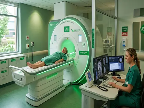
Magnetic Resonance Imaging for Detailed Diagnostic Assessment
The MRI Service at AS Medical Complex provides detailed diagnostic imaging for selected conditions affecting the brain, spine, joints, muscles, soft tissues, abdomen, pelvis, and other internal structures.
Magnetic Resonance Imaging – commonly known as MRI – is a non-invasive imaging technique that uses a powerful magnetic field, radio waves, and computer processing to produce detailed images of structures inside the body.
Unlike conventional X-rays and CT scans, MRI does not use ionizing radiation.
MRI is particularly valuable for evaluating many soft-tissue structures, including:
-
Brain
-
Spinal cord
-
Nerves
-
Intervertebral discs
-
Ligaments
-
Tendons
-
Muscles
-
Cartilage
-
Bone marrow
-
Abdominal organs
-
Pelvic organs
-
Soft-tissue masses
-
Selected blood vessels
-
Other internal structures
MRI can be used to help:
-
Investigate symptoms
-
Diagnose selected diseases
-
Evaluate injuries
-
Assess abnormal findings from another scan
-
Plan treatment
-
Monitor known disease
-
Evaluate selected postoperative conditions
-
Monitor treatment response
At AS Medical Complex, the appropriate MRI examination should be selected according to the patient’s symptoms, medical history, suspected diagnosis, previous investigations, MRI safety screening, and specialist or radiologist recommendation.
Our MRI Services
Depending on clinical indication, available MRI protocols, and specialist recommendation, MRI services at AS Medical Complex may include:
-
MRI Brain
-
MRI Head
-
MRI Pituitary where clinically indicated
-
MRI Orbit where clinically indicated
-
MRI Internal Auditory Canals where available
-
MRI Cervical Spine
-
MRI Thoracic Spine
-
MRI Lumbar Spine
-
MRI Lumbosacral Spine
-
MRI Knee
-
MRI Shoulder
-
MRI Hip
-
MRI Ankle
-
MRI Foot
-
MRI Elbow
-
MRI Wrist
-
MRI Hand
-
Musculoskeletal MRI
-
MRI of muscles and soft tissues
-
MRI Abdomen
-
MRI Pelvis
-
MRI Liver where clinically indicated
-
MRI Pancreas where clinically indicated
-
MRI Kidneys where clinically indicated
-
MRI female pelvis
-
MRI male pelvis
-
MRI prostate where available
-
MRI for selected tumors
-
MRI for selected neurological conditions
-
MRI for selected orthopedic conditions
-
MRI for selected sports injuries
-
MRI without contrast
-
MRI with contrast where clinically indicated
-
Follow-up MRI
-
Selected postoperative MRI
-
MR Angiography – MRA where available
-
MR Cholangiopancreatography – MRCP where available
-
Pediatric MRI where clinically appropriate and available
-
Specialist MRI review
-
Referral for specialized MRI protocols where required
Advanced protocols should only be advertised as onsite services after confirming the equipment, software, contrast capability, coils, and reporting expertise available at AS Medical Complex.
What Is an MRI?
MRI is a diagnostic imaging method that creates highly detailed images of internal body structures.
During an MRI examination, the patient is positioned within a strong magnetic field.
Radiofrequency energy is used to produce signals from tissues within the body.
The MRI system detects these signals and a computer converts them into detailed images.
MRI can produce images in several different planes without physically repositioning the body for every view.
This allows radiologists to evaluate structures from multiple perspectives.
MRI Does Not Use X-Ray Radiation
One important feature of MRI is that it does not use ionizing radiation.
This differs from imaging methods such as:
-
X-ray
-
CT Scan
-
Fluoroscopy
MRI instead uses magnetic fields and radiofrequency energy.
This can make MRI particularly useful when detailed soft-tissue imaging is required.
However, the absence of ionizing radiation does not mean that MRI should automatically be selected instead of every other scan.
Different imaging methods answer different clinical questions.
MRI vs CT Scan
MRI and CT are both important diagnostic imaging techniques, but they work differently.
MRI
MRI is particularly useful for detailed assessment of many:
-
Brain abnormalities
-
Spinal cord abnormalities
-
Nerves
-
Muscles
-
Ligaments
-
Tendons
-
Joint structures
-
Soft tissues
-
Bone marrow
-
Selected abdominal and pelvic conditions
CT Scan
CT is often particularly useful for:
-
Acute trauma
-
Many emergency conditions
-
Lung imaging
-
Certain fractures
-
Acute bleeding
-
Selected abdominal emergencies
-
Rapid assessment of critically ill patients
CT examinations are generally much faster than MRI.
The doctor or radiologist determines which investigation is more appropriate for the clinical problem.
MRI vs X-Ray
X-rays are commonly used for rapid assessment of:
-
Fractures
-
Chest conditions
-
Joint alignment
-
Certain bone abnormalities
MRI provides much more detailed information about many soft tissues.
For example, a knee X-ray may show:
-
Bone
-
Joint space
-
Alignment
while MRI may provide additional information about:
-
Menisci
-
Ligaments
-
Tendons
-
Cartilage
-
Bone marrow
-
Other soft tissues
The tests therefore complement rather than simply replace one another.
MRI vs Ultrasound
Ultrasound and MRI are both non-ionizing imaging methods.
Ultrasound may be particularly useful for:
-
Pregnancy
-
Gallbladder
-
Pelvic assessment
-
Thyroid
-
Superficial lumps
-
Blood-flow studies
-
Other selected conditions
MRI may provide more detailed evaluation of:
-
Brain
-
Spine
-
Deep soft tissues
-
Certain joints
-
Complex pelvic conditions
-
Selected abdominal abnormalities
The most appropriate test depends on the clinical question.
MRI Brain
MRI is one of the most important imaging techniques for detailed assessment of the brain.
A brain MRI may be recommended in selected patients with:
-
Persistent neurological symptoms
-
Suspected brain tumor
-
Seizures
-
Suspected multiple sclerosis
-
Selected stroke-related conditions
-
Brain inflammation
-
Brain infection
-
Certain congenital abnormalities
-
Pituitary abnormalities
-
Cranial-nerve disorders
-
Abnormal CT findings
-
Unexplained neurological deterioration
-
Follow-up of previously identified brain lesions
MRI provides detailed images of brain tissue and related structures.
Headache & MRI
Headache is common and does not automatically require MRI.
Many headaches can be diagnosed and treated based on:
-
Medical history
-
Neurological examination
-
Headache pattern
-
Other clinical information
MRI may be considered when there are concerning features such as:
-
New neurological abnormalities
-
Significant change in headache pattern
-
New severe headache in selected high-risk patients
-
Suspicion of structural brain disease
-
Other clinically concerning features
Patients should be assessed by an appropriate doctor rather than requesting MRI solely because they have headache.
MRI & Stroke
MRI can help identify selected abnormalities related to stroke and other cerebrovascular conditions.
However, acute stroke is a medical emergency.
Symptoms may include:
-
Sudden facial weakness
-
Sudden weakness of an arm or leg
-
Sudden difficulty speaking
-
Sudden difficulty understanding speech
-
Sudden loss of balance
-
Sudden visual disturbance
-
Sudden severe neurological deterioration
A patient with possible acute stroke should go to Emergency care immediately.
Emergency imaging may involve:
-
CT
-
CT angiography
-
MRI
-
MR angiography
-
Other imaging
depending on the clinical situation and available emergency pathway.
A patient should not wait for a routine outpatient MRI appointment when stroke is suspected.
MRI for Seizures
MRI may be useful in selected patients with:
-
First seizure
-
Recurrent seizures
-
Focal seizures
-
Abnormal neurological examination
-
Suspected structural brain abnormality
-
Difficult-to-control epilepsy
MRI may help identify structural abnormalities that can contribute to seizures.
However, seizure assessment may also require:
-
Neurology consultation
-
EEG
-
Blood tests
-
Medication review
-
Other investigations
MRI alone does not diagnose every cause of seizure.
MRI for Multiple Sclerosis
MRI plays an important role in the assessment and monitoring of selected patients with suspected or established multiple sclerosis and other demyelinating conditions.
Imaging may involve:
-
Brain
-
Cervical spinal cord
-
Thoracic spinal cord
depending on symptoms and specialist recommendation.
Contrast may sometimes be used to help evaluate lesion activity.
MRI findings must be interpreted together with:
-
Symptoms
-
Neurological examination
-
Disease history
-
Other diagnostic criteria
-
Other investigations
MRI Pituitary
A specialized pituitary MRI may be requested when there is concern about the pituitary gland.
Possible reasons include:
-
Hormonal abnormalities
-
Suspected pituitary adenoma
-
Abnormal prolactin or other hormone results
-
Visual symptoms associated with suspected pituitary disease
-
Other endocrine concerns
Evaluation may involve coordination with:
-
Endocrinology
-
Neurology
-
Neurosurgery
-
Ophthalmology
depending on the findings.
MRI Orbit
MRI of the orbit may be considered for selected abnormalities involving:
-
Optic nerves
-
Eye muscles
-
Soft tissues behind the eyes
-
Selected orbital tumors
-
Inflammatory conditions
-
Other orbital abnormalities
The appropriate protocol depends on the suspected diagnosis.
MRI Internal Auditory Canals
Specialized MRI can be used in selected patients when doctors need detailed assessment of structures related to:
-
Hearing nerves
-
Balance nerves
-
Internal auditory canals
-
Selected cranial nerves
Possible reasons may include:
-
Asymmetric hearing loss
-
Certain neurological symptoms
-
Suspected vestibular schwannoma
-
Other specialist indications
ENT, Neurology, or Audiology assessment may also be required.
MRI Spine
MRI is particularly useful for examining structures within and around the spine.
These include:
-
Spinal cord
-
Nerve roots
-
Intervertebral discs
-
Spinal canal
-
Ligaments
-
Bone marrow
-
Surrounding soft tissues
Spinal MRI may be performed for:
-
Cervical spine
-
Thoracic spine
-
Lumbar spine
-
Lumbosacral region
depending on symptoms.
MRI Cervical Spine
Cervical-spine MRI examines the neck portion of the spine.
It may be requested for selected patients with:
-
Neck pain associated with neurological symptoms
-
Arm pain
-
Arm numbness
-
Arm weakness
-
Suspected cervical-disc disease
-
Spinal stenosis
-
Suspected spinal-cord compression
-
Selected trauma
-
Suspected infection
-
Suspected tumor
-
Postoperative assessment
MRI can help evaluate:
-
Discs
-
Spinal cord
-
Nerve roots
-
Spinal canal
-
Other surrounding structures
MRI Thoracic Spine
Thoracic-spine MRI may be considered for selected patients with:
-
Thoracic spinal pain
-
Neurological abnormalities
-
Suspected cord compression
-
Tumor
-
Infection
-
Fracture
-
Demyelinating disease
-
Other spinal conditions
Because the spinal cord extends through the thoracic region, neurological symptoms can sometimes require detailed thoracic imaging.
MRI Lumbar Spine
Lumbar-spine MRI is commonly used for selected lower-back conditions.
Possible indications include:
-
Sciatica
-
Lumbar-disc herniation
-
Persistent neurological symptoms
-
Leg weakness
-
Significant numbness
-
Spinal stenosis
-
Nerve-root compression
-
Suspected infection
-
Suspected tumor
-
Significant trauma
-
Postoperative assessment
Routine MRI is not required for every episode of uncomplicated low-back pain.
Clinical assessment should guide imaging.
Disc Herniation
Intervertebral discs can develop:
-
Bulging
-
Protrusion
-
Extrusion
-
Degenerative changes
MRI can show:
-
Disc position
-
Relationship with nerves
-
Spinal canal
-
Associated degenerative changes
However, an abnormal MRI does not automatically mean surgery is required.
Some disc changes are also seen in people without significant symptoms.
Treatment decisions should consider:
-
Pain
-
Weakness
-
Numbness
-
Neurological examination
-
Functional limitation
-
MRI findings
-
Response to conservative treatment
Sciatica
Sciatica commonly refers to pain traveling from the lower back or buttock into the leg because of irritation or compression involving a nerve root.
Symptoms may include:
-
Leg pain
-
Tingling
-
Numbness
-
Weakness
MRI may be useful in selected patients, particularly when:
-
Neurological symptoms are significant
-
Symptoms persist
-
Surgery is being considered
-
Serious underlying disease is suspected
Many patients with uncomplicated sciatica do not require immediate MRI.
Spinal Stenosis
Spinal stenosis refers to narrowing within the spinal canal or pathways through which nerves travel.
MRI may help determine:
-
Location
-
Severity
-
Disc contribution
-
Joint changes
-
Nerve involvement
-
Spinal-cord involvement
Treatment may involve:
-
Physiotherapy
-
Medication
-
Pain-management strategies
-
Ortho & Spine Surgery
-
Neurosurgery
depending on severity.
Spinal Cord Compression
Spinal-cord compression can become serious.
Symptoms may include:
-
Progressive weakness
-
Difficulty walking
-
Loss of balance
-
Numbness
-
Changes in hand coordination
-
Bladder or bowel problems
Patients with rapidly progressive symptoms require urgent medical assessment.
Cauda Equina Syndrome
Severe compression of nerves at the lower end of the spinal canal can produce cauda equina syndrome.
Warning symptoms can include:
-
New urinary retention
-
New urinary incontinence
-
New loss of bowel control
-
Saddle-region numbness
-
Severe bilateral leg symptoms
-
Progressive leg weakness
This is a medical emergency.
Urgent clinical and MRI assessment may be required.
A patient with suspected cauda equina syndrome should not wait for a routine MRI booking.
Musculoskeletal MRI
MRI provides detailed assessment of many musculoskeletal structures.
These include:
-
Ligaments
-
Tendons
-
Muscles
-
Cartilage
-
Bone marrow
-
Joint structures
-
Soft tissues
MRI is particularly valuable because these structures may not be well visualized on routine X-rays.
MRI Knee
Knee MRI may help evaluate:
-
ACL
-
PCL
-
Menisci
-
Collateral ligaments
-
Tendons
-
Cartilage
-
Bone marrow
-
Joint fluid
-
Other knee structures
Possible reasons include:
-
Sports injury
-
Twisting injury
-
Suspected ligament tear
-
Suspected meniscal tear
-
Persistent symptoms after trauma
-
Selected preoperative planning
Not every patient with knee pain requires MRI.
Clinical examination and X-rays may sometimes provide enough information initially.
ACL Injury
The Anterior Cruciate Ligament – ACL – may be injured during:
-
Sports
-
Sudden twisting
-
Direction changes
-
Falls
-
Other trauma
MRI can help assess:
-
ACL integrity
-
Other ligament injuries
-
Meniscal injuries
-
Bone bruising
-
Cartilage
-
Joint fluid
Treatment depends on:
-
Degree of instability
-
Associated injuries
-
Age
-
Activity level
-
Sport
-
Patient goals
not only the MRI report.
Meniscus Tear
The knee contains medial and lateral menisci.
MRI can help identify many meniscal tears.
However, some meniscal abnormalities are degenerative and may be seen in patients who do not have significant symptoms.
Orthopedic assessment is therefore important before deciding on surgery.
MRI Shoulder
MRI can provide detailed imaging of:
-
Rotator-cuff tendons
-
Muscles
-
Labrum
-
Ligaments
-
Cartilage
-
Bone marrow
-
Other shoulder structures
MRI may be considered for:
-
Rotator-cuff injury
-
Shoulder instability
-
Labral injury
-
Persistent shoulder symptoms
-
Selected trauma
-
Other soft-tissue conditions
Rotator Cuff
The rotator cuff is a group of muscles and tendons involved in shoulder movement and stability.
MRI can help identify:
-
Tendon degeneration
-
Partial tears
-
Full-thickness tears
-
Muscle changes
-
Associated abnormalities
Treatment may include:
-
Physiotherapy
-
Medication
-
Activity modification
-
Selected injections
-
Surgery in selected cases
depending on the complete clinical assessment.
MRI Hip
Hip MRI may be useful for selected evaluation of:
-
Soft-tissue injuries
-
Tendon abnormalities
-
Cartilage
-
Bone marrow
-
Selected stress injuries
-
Certain fractures not clearly visible on initial imaging
-
Other hip conditions
The appropriate imaging technique depends on symptoms and examination.
MRI Ankle & Foot
MRI may help assess selected:
-
Ligament injuries
-
Tendon injuries
-
Cartilage abnormalities
-
Stress injuries
-
Bone-marrow abnormalities
-
Soft-tissue masses
-
Other ankle and foot conditions
Sports injuries may sometimes require Orthopedic or Physiotherapy assessment before MRI is ordered.
MRI Wrist & Hand
MRI may be useful for selected conditions involving:
-
Ligaments
-
Tendons
-
Soft tissues
-
Nerves
-
Bone marrow
-
Cartilage
-
Other structures
The scan should be targeted to the exact anatomical region requiring evaluation.
MRI Elbow
Elbow MRI may provide detailed assessment of selected:
-
Tendon injuries
-
Ligament injuries
-
Muscle abnormalities
-
Joint abnormalities
-
Bone-marrow changes
The clinical indication should determine whether MRI is preferable to X-ray, ultrasound, or another investigation.
Sports Injury MRI
MRI is commonly associated with sports-medicine imaging because it provides detailed visualization of:
-
Ligaments
-
Tendons
-
Muscles
-
Cartilage
-
Bone marrow
However, not every sports injury requires MRI.
Initial assessment may involve:
-
Medical examination
-
Orthopedic assessment
-
X-ray
-
Physiotherapy assessment
-
Other investigations
MRI is most useful when its findings are expected to affect treatment planning.
Soft-Tissue MRI
MRI can help assess selected soft-tissue abnormalities including:
-
Muscle injuries
-
Deep soft-tissue masses
-
Certain tumors
-
Selected infections
-
Other abnormal tissue
If a mass is detected, further evaluation may require:
-
Ultrasound
-
CT
-
MRI with contrast
-
Biopsy
-
Pathology
-
Surgical assessment
MRI cannot always determine whether a mass is benign or malignant without tissue diagnosis.
MRI Abdomen
MRI of the abdomen may provide detailed images of selected abdominal organs.
Depending on the indication, these may include:
-
Liver
-
Pancreas
-
Kidneys
-
Biliary system
-
Other abdominal structures
Abdominal MRI may be used for:
-
Characterization of abnormalities found on ultrasound or CT
-
Selected liver lesions
-
Selected pancreatic abnormalities
-
Selected kidney abnormalities
-
Selected tumors
-
Other specialist indications
MRI Liver
MRI may be useful for characterizing selected liver lesions.
The examination may provide additional information about:
-
Size
-
Location
-
Tissue characteristics
-
Relationship with surrounding structures
-
Enhancement after contrast when used
A liver lesion does not automatically mean cancer.
Possible liver abnormalities include:
-
Cysts
-
Hemangiomas
-
Other benign lesions
-
Inflammatory abnormalities
-
Malignant lesions
Further specialist assessment may be required.
MRI Pancreas
Selected pancreatic abnormalities may require MRI or MRCP.
MRI may be used to assess:
-
Pancreatic lesions
-
Cysts
-
Selected inflammatory changes
-
Pancreatic and biliary anatomy
-
Other abnormalities
Gastroenterology or General Surgery assessment may be appropriate depending on findings.
MRCP
Magnetic Resonance Cholangiopancreatography – MRCP is a specialized MRI technique used to obtain images of structures such as the:
-
Bile ducts
-
Gallbladder
-
Pancreatic duct
-
Pancreas
It may be used for selected patients with suspected:
-
Bile-duct stones
-
Biliary obstruction
-
Pancreatic or biliary abnormalities
-
Other appropriate conditions
MRCP availability and protocol at AS Medical Complex should be confirmed before booking.
MRI Pelvis
Pelvic MRI may provide detailed assessment of structures within the pelvis.
Depending on sex and clinical indication, these can include:
-
Uterus
-
Cervix
-
Ovaries
-
Bladder
-
Prostate
-
Rectum
-
Pelvic soft tissues
-
Other structures
MRI pelvis may be requested by:
-
Gynecology
-
Urology
-
General Surgery
-
Oncology
-
Other specialists
MRI Female Pelvis
MRI may be considered for selected gynecological conditions involving:
-
Fibroids
-
Adenomyosis
-
Endometriosis
-
Ovarian or pelvic masses
-
Cervical abnormalities
-
Other complex pelvic conditions
Ultrasound is often the first imaging investigation for many gynecological problems.
MRI may provide additional information when clinically required.
Fibroids & MRI
Fibroids are benign muscular growths arising from the uterus.
Many fibroids can be evaluated with ultrasound.
MRI may be helpful when:
-
There are multiple large fibroids
-
Anatomy is complex
-
Treatment planning requires additional detail
-
Ultrasound findings are uncertain
-
Another pelvic condition is suspected
MRI does not automatically need to be performed for every patient with fibroids.
Endometriosis
MRI may help assess selected patients with suspected deep or complex endometriosis.
However, diagnosis and treatment planning also depend on:
-
Symptoms
-
Gynecological assessment
-
Ultrasound
-
Surgical history
-
Other investigations
MRI is one part of the overall evaluation.
MRI Prostate
Multiparametric prostate MRI may be used in selected patients for:
-
Assessment of suspicious prostate abnormalities
-
Evaluation after abnormal prostate-related testing
-
Biopsy planning
-
Selected cancer assessment
-
Treatment planning
This requires a specialized MRI protocol.
Availability of prostate MRI at AS Medical Complex should therefore be confirmed before it is advertised as a routine onsite service.
MRI for Tumors
MRI can provide detailed information about selected tumors involving:
-
Brain
-
Spine
-
Soft tissues
-
Musculoskeletal structures
-
Abdomen
-
Pelvis
-
Other areas
MRI may help determine:
-
Size
-
Location
-
Relationship to surrounding tissues
-
Certain tissue characteristics
-
Changes over time
-
Response to treatment
However, MRI does not replace biopsy when tissue diagnosis is required.
MRI & Cancer Diagnosis
A suspicious MRI finding does not automatically confirm cancer.
Depending on the lesion, further assessment may include:
-
Clinical examination
-
Laboratory investigations
-
CT
-
Ultrasound
-
Endoscopy
-
Colonoscopy
-
Biopsy
-
Histopathology
-
Oncology assessment
MRI contributes to diagnosis and treatment planning but should be interpreted within the full clinical pathway.
MRI With Contrast
Some MRI examinations are performed with an intravenous contrast agent.
The most commonly used MRI contrast agents are based on gadolinium.
Contrast may help the radiologist obtain additional information about:
-
Blood vessels
-
Inflammation
-
Tumors
-
Infection
-
Blood-brain barrier abnormalities
-
Tissue enhancement
-
Other selected conditions
Not every MRI examination requires contrast.
The decision depends on the clinical question and MRI protocol.
MRI Without Contrast
Many MRI examinations can be performed without intravenous contrast.
Examples may include selected:
-
Spine MRI
-
Knee MRI
-
Joint MRI
-
Other musculoskeletal studies
depending on the clinical indication.
Patients should not assume that a contrast study is automatically “better.”
Contrast should be used when it provides meaningful additional diagnostic information.
Gadolinium Contrast Safety
Gadolinium-based contrast agents are generally well tolerated when appropriately used, but they are not completely risk-free.
Possible issues include:
-
Allergic or hypersensitivity reactions
-
Kidney-related considerations in selected patients
-
Other uncommon complications
FDA notes that gadolinium-based contrast agents are administered intravenously for selected MRI examinations and that their risks should be considered according to the patient’s clinical situation. (fda.gov)
Kidney Disease & MRI Contrast
Patients should tell the imaging team if they have:
-
Chronic Kidney Disease
-
Kidney failure
-
Dialysis
-
Recent acute kidney injury
-
Previous significant kidney problems
Severe kidney dysfunction can affect decisions regarding certain gadolinium-based contrast agents.
The Radiology team may need to review:
-
Kidney function
-
Recent creatinine
-
eGFR
-
Dialysis status
-
Clinical necessity of contrast
before proceeding.
The exact requirement depends on the contrast agent and patient risk.
Contrast Allergies
Patients should inform the Radiology team if they have previously experienced a reaction to MRI contrast.
Possible reactions can include:
-
Rash
-
Hives
-
Swelling
-
Breathing difficulty
-
Other symptoms
A previous reaction does not automatically mean MRI can never be performed again.
The radiologist determines the safest approach.
MRI & Pregnancy
Patients should always tell the doctor and MRI team if they:
-
Are pregnant
-
Think they may be pregnant
MRI does not use ionizing radiation.
Current radiology safety information states that no established harmful effects from routine non-contrast MRI have been demonstrated in pregnancy, but non-urgent examinations may sometimes be postponed depending on gestational stage and clinical need.
The decision should balance:
-
Clinical necessity
-
Timing
-
Alternative investigations
-
Benefits of diagnosis
-
Potential uncertainties
Gadolinium Contrast During Pregnancy
Gadolinium contrast is generally avoided during pregnancy unless the expected diagnostic benefit clearly justifies its use.
RadiologyInfo advises that gadolinium contrast is usually avoided in pregnancy except in specific circumstances where it is considered medically necessary.
The radiologist and treating specialist should make this decision.
MRI Safety Screening
MRI safety screening is essential because the MRI magnet is extremely powerful.
Before entering the MRI environment, patients may be asked detailed questions regarding:
-
Pacemaker
-
Implantable cardiac device
-
Cochlear implant
-
Brain aneurysm clips
-
Vascular coils
-
Neurostimulator
-
Insulin pump
-
Implanted infusion pump
-
Artificial joint
-
Orthopedic hardware
-
Previous surgery
-
Metal fragments
-
Previous gunshot injury
-
Eye injury involving metal
-
Other implants or devices
Patients should answer these questions accurately.
Pacemakers & MRI
Modern cardiac devices vary substantially in MRI compatibility.
Some are:
-
MR Safe
-
MR Conditional
-
Not suitable for MRI
-
Unknown in MRI safety status
MRI should never be performed simply because a patient says that the device “should be okay.”
FDA guidance states that implanted devices should be positively identified as MR Safe or MR Conditional before MRI, and MR Conditional devices must only be scanned under their specified conditions.
Patients should bring:
-
Implant card
-
Device information
-
Manufacturer details
-
Previous medical documentation
where available.
Cochlear Implants
Some cochlear implants require special precautions or may not be suitable for MRI under certain conditions.
Patients with a cochlear implant must inform the MRI team before entering the scanner area.
The exact implant model should be identified whenever possible.
Aneurysm Clips & Coils
Certain clips used in previous brain aneurysm surgery can create MRI safety concerns.
Patients should tell the imaging team about:
-
Previous aneurysm surgery
-
Brain clips
-
Vascular coils
-
Other neurovascular implants
The implant must be appropriately evaluated before MRI.
Orthopedic Implants
Many modern orthopedic implants can be safely scanned under appropriate conditions.
Examples may include:
-
Joint replacements
-
Plates
-
Screws
-
Rods
-
Other surgical hardware
However, every implant should still be disclosed during MRI screening.
Metal can also produce image artifacts and reduce diagnostic quality around the implant.
Metal Fragments
Patients should inform the MRI team if they have a history of:
-
Metalworking
-
Welding
-
Grinding metal
-
Eye injury involving metal
-
Gunshot wound
-
Shrapnel
-
Other retained metallic fragments
Certain metal fragments can pose a safety risk.
Additional evaluation or X-ray may sometimes be necessary before MRI.
Jewelry & Metal Objects
Before MRI, patients may need to remove items such as:
-
Jewelry
-
Watches
-
Earrings
-
Chains
-
Rings
-
Hairpins
-
Metal piercings
-
Removable dental items
-
Hearing aids
-
Wallets
-
Keys
-
Coins
-
Mobile phones
-
Other metallic or electronic objects
The exact requirements depend on MRI safety procedures.
Electronic Devices
The MRI magnetic field can damage or interfere with electronic devices.
Items such as:
-
Mobile phones
-
Smart watches
-
Bank cards
-
Hearing devices
-
Other electronics
should not be taken into restricted MRI areas unless specifically approved as MRI-compatible.
What Should You Wear for MRI?
Patients may be asked to change into a hospital gown.
Clothing containing:
-
Metal fasteners
-
Zippers
-
Hooks
-
Metallic fibers
-
Other metal components
may not be suitable within the scanner.
The MRI team will provide instructions.
Preparing for MRI
Preparation depends on the type of examination.
Patients may be instructed regarding:
-
Food
-
Drink
-
Medication
-
Contrast
-
Kidney-function testing
-
Clothing
-
Metal objects
-
Previous medical reports
Unless specifically told otherwise, many routine MRI examinations do not require major changes in normal medication or food intake.
RadiologyInfo advises patients to follow the specific instructions supplied for their MRI because preparation varies by examination.
Fasting Before MRI
Not every MRI requires fasting.
Fasting may be requested for:
-
Selected abdominal MRI examinations
-
MRCP
-
Procedures involving sedation
-
Certain contrast protocols
-
Other specific examinations
Patients should follow the instructions provided for their exact MRI study.
Medicines Before MRI
Patients should usually continue regular medicines unless specifically instructed otherwise.
Tell the imaging team about all relevant medical conditions and medication.
Patients should not independently stop:
-
Blood-pressure medicine
-
Heart medicine
-
Diabetes medicine
-
Other prescribed treatment
without appropriate medical instructions.
Diabetes & MRI
Patients with diabetes should inform the imaging team, particularly if:
-
Fasting is required
-
Sedation is planned
-
Contrast is planned
-
Kidney disease is present
Meal and medication timing may require individualized instructions.
Claustrophobia
Some patients feel anxious or uncomfortable inside the MRI scanner.
This is known as claustrophobia.
Possible strategies include:
-
Explaining the procedure beforehand
-
Communication with the technologist
-
Relaxation techniques
-
Appropriate positioning
-
Mild sedation in selected patients
RadiologyInfo advises patients with significant claustrophobia or anxiety to discuss possible sedation with their doctor before the appointment.
Sedation for MRI
Selected patients may require sedation because of:
-
Severe claustrophobia
-
Inability to remain still
-
Certain neurological conditions
-
Pediatric age
-
Severe anxiety
-
Other clinical factors
Sedation requires additional safety considerations.
Depending on the level of sedation, patients may need:
-
Fasting
-
Pre-procedure assessment
-
Monitoring
-
Anesthesia involvement
-
Someone to take them home
Patients receiving sedating medication should not drive themselves afterward.
Availability of sedation-supported MRI at AS Medical Complex should be confirmed before booking.
MRI for Children
Children may require MRI for selected:
-
Neurological conditions
-
Brain abnormalities
-
Spine problems
-
Musculoskeletal conditions
-
Tumors
-
Other conditions
MRI has the advantage of not using ionizing radiation.
However, children must remain very still during image acquisition.
Younger children may sometimes require:
-
Preparation
-
Reassurance
-
Sedation
-
Anesthesia
depending on age and ability to cooperate.
Pediatric sedation or anesthesia should only be offered where appropriate facilities and trained personnel are available.
What Happens During an MRI?
Before the scan:
-
The examination is confirmed.
-
Safety screening is completed.
-
Metal objects are removed.
-
An IV line may be inserted if contrast is required.
-
The patient is positioned on the MRI table.
-
A specialized imaging coil may be placed around the body part being scanned.
The table then moves into the MRI scanner.
During scanning, the patient must remain as still as possible.
MRI Noise
MRI scanners produce loud repetitive sounds during image acquisition.
These may include:
-
Knocking
-
Tapping
-
Buzzing
-
Thumping
Patients are generally provided with hearing protection according to facility procedures.
The noise is produced by normal operation of the MRI system.
Staying Still During MRI
Movement can significantly reduce MRI image quality.
Patients should remain still during each imaging sequence.
If the scan becomes blurred because of movement, some sequences may need to be repeated.
Children and patients with pain or neurological conditions may find this more difficult.
Appropriate positioning and communication can help.
Communication During MRI
Although the technologist usually operates the scanner from a separate control area, the patient can normally be monitored and communicated with during the examination.
Patients should immediately tell the technologist if they experience:
-
Severe pain
-
Breathing difficulty
-
Panic
-
Significant discomfort
-
Any unexpected problem
How Long Does an MRI Take?
MRI examination time varies substantially.
It depends on:
-
Body area
-
Number of sequences
-
Whether contrast is required
-
Patient movement
-
Complexity of the examination
-
Specialized protocols
Some MRI studies are relatively short, while complex examinations may take considerably longer.
AS Medical Complex should provide an estimated duration according to the specific scan rather than promising one universal MRI time.
After MRI Without Sedation
After a routine MRI without sedation, most patients can usually return to normal activities unless their doctor has provided other instructions.
If IV contrast was used, the patient may receive additional instructions according to the examination and clinical circumstances.
After MRI With Sedation
Patients who receive sedation may require:
-
Recovery monitoring
-
Assistance traveling home
-
Temporary restriction from driving
-
Other instructions
The exact precautions depend on the medication used.
MRI Results
MRI images are interpreted by a radiologist or another appropriately qualified imaging specialist.
The report may describe:
-
Normal findings
-
Abnormal findings
-
Location of abnormalities
-
Size
-
Tissue characteristics
-
Relationship to surrounding structures
-
Comparison with previous imaging
-
Recommendations for additional imaging or specialist assessment
The report should be interpreted together with the patient’s clinical condition.
MRI Images vs MRI Report
The MRI examination produces a large collection of images.
The radiologist evaluates these images and prepares a report.
Patients may receive:
-
Images
-
Digital access
-
Report
-
Other documentation
depending on hospital systems.
The written report summarizes the radiologist’s interpretation.
Abnormal MRI Does Not Always Mean Serious Disease
MRI is very sensitive and may identify changes that are:
-
Mild
-
Degenerative
-
Age-related
-
Incidental
-
Unrelated to the patient’s symptoms
For example, an MRI may show spinal-disc changes that are not actually responsible for the patient’s pain.
Treatment decisions should therefore be based on both:
-
Clinical symptoms
-
Imaging findings
Incidental Findings
An incidental finding is an abnormality discovered unexpectedly while imaging another problem.
Depending on the finding, the doctor may recommend:
-
No further treatment
-
Follow-up imaging
-
Another type of scan
-
Specialist consultation
-
Additional investigation
Not every incidental finding is dangerous.
Comparing With Previous MRI
Previous imaging can be valuable.
Patients should bring or provide earlier:
-
MRI
-
CT
-
X-ray
-
Ultrasound
-
Reports
when available.
Comparison may help determine whether an abnormality is:
-
New
-
Stable
-
Improving
-
Progressing
MRI After Surgery
MRI may be useful in selected postoperative situations.
Possible reasons include:
-
Persistent symptoms
-
Suspected complication
-
Recurrent disc disease
-
Tumor follow-up
-
Other surgical concerns
However, metal implants can sometimes create image artifacts.
The radiologist may need specific imaging techniques.
Patients should provide information about:
-
Surgery
-
Implant type
-
Date
-
Previous operative records
MRI & Orthopedics
The Orthopedics Department may request MRI for selected:
-
Ligament injuries
-
Tendon injuries
-
Meniscal injuries
-
Joint problems
-
Bone-marrow abnormalities
-
Sports injuries
-
Soft-tissue conditions
The orthopedic specialist uses imaging together with examination to determine treatment.
MRI & Ortho & Spine Surgery
Spine specialists may use MRI to assess:
-
Disc herniation
-
Nerve compression
-
Spinal stenosis
-
Spinal-cord abnormalities
-
Infection
-
Tumor
-
Postoperative problems
Surgery is not recommended simply because an MRI shows a disc abnormality.
The clinical significance of the finding must be established.
MRI & Neurosurgery
Neurosurgery may use MRI for selected conditions involving:
-
Brain tumors
-
Spinal tumors
-
Hydrocephalus
-
Spinal-cord compression
-
Disc disease
-
Other neurological structural abnormalities
The MRI can support diagnosis and surgical planning when required.
MRI & Neurology
Neurology may use MRI in the assessment of:
-
Seizures
-
Multiple sclerosis
-
Stroke-related disease
-
Certain headaches
-
Movement disorders
-
Dementia-related conditions in selected cases
-
Other neurological disorders
MRI findings are interpreted alongside neurological examination.
MRI & Gynecology
Gynecology may request MRI for selected:
-
Fibroids
-
Adenomyosis
-
Endometriosis
-
Pelvic masses
-
Complex ovarian abnormalities
-
Other reproductive-system conditions
Ultrasound remains an important first-line imaging method for many gynecological conditions.
MRI & Urology
Urology may use selected MRI protocols for:
-
Prostate assessment
-
Pelvic abnormalities
-
Selected kidney or urinary conditions
-
Other specialist indications
Some examinations require specialized protocols and reporting expertise.
MRI & Gastroenterology
Gastroenterology may use abdominal MRI or MRCP in selected patients with:
-
Liver abnormalities
-
Pancreatic disease
-
Biliary disease
-
Other gastrointestinal conditions
The appropriate investigation may be MRI, CT, ultrasound, endoscopy, or another test depending on the condition.
MRI & Oncology
MRI can play an important role in selected cancer pathways.
It may be used for:
-
Tumor characterization
-
Local staging
-
Treatment planning
-
Treatment response
-
Follow-up
depending on cancer type.
The Oncology team may coordinate imaging with:
-
Radiology
-
Surgery
-
Pathology
-
Other departments
MRI & Radiology
MRI is part of the wider Radiology Service at AS Medical Complex.
Radiologists help determine:
-
Appropriate MRI protocol
-
Need for contrast
-
Important safety considerations
-
Image interpretation
-
Need for further imaging
Clinical information from the referring doctor is important because it helps the radiologist tailor the examination to the medical question.
MRI Limitations
MRI is an extremely useful imaging technique, but it also has limitations.
These may include:
-
Longer examination time than CT
-
Sensitivity to patient movement
-
Claustrophobia
-
Safety considerations with certain implants
-
Difficulty imaging selected patients
-
Metal-related artifacts
-
Limited suitability for some emergency situations
-
Need for sedation in selected patients
-
Higher complexity for certain protocols
MRI should therefore be selected when its diagnostic advantages are appropriate for the clinical problem.
MRI Is Not Always the Best Emergency Scan
MRI can provide excellent detail but takes longer than many CT examinations.
In emergencies involving:
-
Major trauma
-
Suspected acute intracranial bleeding
-
Certain acute abdominal conditions
-
Critically unstable patients
CT may sometimes be preferred because of speed and accessibility.
The Emergency and Radiology teams determine the appropriate imaging pathway.
When Should You Consider an MRI?
You may require MRI assessment if your doctor needs detailed imaging for:
-
Neurological symptoms
-
Suspected brain disease
-
Persistent spine symptoms with neurological features
-
Nerve compression
-
Joint injury
-
Ligament or tendon injury
-
Sports injury
-
Soft-tissue abnormality
-
Pelvic disease
-
Selected abdominal conditions
-
Suspected tumor
-
Follow-up of known disease
-
Abnormal findings on another imaging study
-
Postoperative evaluation
MRI should ideally answer a specific clinical question.
When Should MRI Safety Be Reviewed Before Booking?
Contact the MRI or Radiology team before the examination if you have:
-
Pacemaker
-
Implantable defibrillator
-
Cochlear implant
-
Brain aneurysm clip
-
Vascular coil
-
Neurostimulator
-
Implanted pump
-
Other electronic implant
-
Retained metal fragment
-
Previous metal injury to the eye
-
Significant surgical hardware
-
Pregnancy
-
Severe kidney disease when contrast may be required
-
Previous MRI contrast reaction
-
Severe claustrophobia
Do not assume an implant is MRI-compatible without proper verification.
FDA recommends that an implanted device be positively identified as MR Safe or MR Conditional and, when conditional, scanned only under the conditions specified for that device.
When Is Emergency Care Needed Instead of Routine MRI?
Do not wait for a routine MRI appointment if you develop:
-
Sudden facial weakness
-
Sudden arm or leg weakness
-
Sudden speech difficulty
-
Loss of consciousness
-
New severe neurological deterioration
-
New paralysis
-
Severe spinal pain with new bladder or bowel dysfunction
-
Saddle-region numbness
-
Major trauma with neurological symptoms
-
Seizure with prolonged loss of consciousness or serious deterioration
-
Other potentially life-threatening symptoms
These situations require urgent clinical assessment.
The Emergency team will determine whether MRI, CT, or another investigation is needed.
Multidisciplinary MRI Care
Depending on the MRI findings, care may be coordinated with:
-
Radiology
-
Neurology
-
Neurosurgery
-
Orthopedics
-
Ortho & Spine Surgery
-
General Surgery
-
Gastroenterology
-
Urology
-
Gynecology
-
Oncology
-
ENT
-
Ophthalmology
-
Endocrinology
-
Physiotherapy
-
Other relevant specialties
MRI is therefore often one stage within a larger diagnostic and treatment pathway.
Follow-Up After MRI
The next step after MRI depends on the findings.
Possible outcomes include:
-
No further investigation
-
Medical treatment
-
Physiotherapy
-
Specialist referral
-
Follow-up imaging
-
Surgery
-
Biopsy
-
Other diagnostic testing
Patients should review significant MRI findings with the doctor responsible for their care rather than relying only on the wording of the report.
Repeat MRI
Repeat MRI may be appropriate for selected patients when:
-
A known disease requires monitoring
-
Symptoms change
-
Treatment response needs assessment
-
Postoperative follow-up is required
-
A previous finding requires surveillance
Repeat imaging should be based on clinical need.
There is usually no benefit in repeatedly performing MRI at arbitrary intervals without a medical indication.
MRI FAQs
What is an MRI?
MRI – Magnetic Resonance Imaging – is a non-invasive imaging technique that uses a powerful magnetic field, radio waves, and computer processing to create detailed images of structures inside the body. It is particularly useful for many soft tissues, including the brain, spinal cord, muscles, ligaments, tendons, and joints, and it does not use ionizing X-ray radiation.
Is MRI safe?
MRI is generally safe when appropriate safety screening is performed. The strong magnetic field means that certain implants, devices, and metal fragments require careful evaluation before the patient enters the MRI environment. Patients should disclose all implants and previous metal injuries. FDA advises that implanted devices should be positively identified as MR Safe or MR Conditional before MRI.
Does every MRI require contrast?
No. Many MRI examinations are performed without contrast. Gadolinium-based contrast may be used when it provides additional diagnostic information about tumors, inflammation, blood vessels, infection, or other selected abnormalities. The radiologist determines whether contrast is necessary for the clinical question.
Can I have an MRI if I have a pacemaker or metal implant?
Possibly, but the exact implant must be identified first. Some devices are MR Safe, some are MR Conditional and can only be scanned under specific conditions, while others may not be suitable for MRI. Bring your implant card or device information whenever possible and never enter the MRI area until the device has been cleared by the MRI team.
Can a pregnant woman have an MRI?
MRI does not use ionizing radiation, and current evidence has not established harmful fetal effects from routine non-contrast MRI. Nevertheless, pregnancy should always be disclosed, and the examination should be performed when medically justified. Gadolinium contrast is generally avoided during pregnancy unless there is a specific clinical reason where the expected benefit outweighs the concern.
What if I am claustrophobic?
Tell the MRI team before your appointment. Explanation, reassurance, communication during the scan, and other measures may help. Patients with significant claustrophobia may sometimes be prescribed a mild sedative. If sedation is used, additional preparation may be necessary and the patient should arrange transportation rather than driving afterward.
How long does an MRI take?
MRI examination time varies depending on the body part, number of imaging sequences, need for contrast, specialized protocols, and the patient’s ability to remain still. Some examinations are relatively short while complex MRI studies take longer. The MRI team should provide an estimated duration for the specific scan rather than applying one fixed time to every examination.
MRI Services at AS Medical Complex
The MRI Service at AS Medical Complex is designed to provide detailed, non-ionizing diagnostic imaging for appropriately selected patients.
From MRI of the brain and spine to joint, musculoskeletal, abdominal, pelvic, soft-tissue, neurological, orthopedic, and selected tumor imaging, our goal is to provide high-quality diagnostic information that supports accurate clinical decision-making.
MRI safety remains an essential part of every examination. Patients should always disclose pacemakers, implanted electronic devices, cochlear implants, aneurysm clips, vascular implants, surgical hardware, retained metal fragments, pregnancy, kidney disease, previous contrast reactions, and severe claustrophobia before entering the MRI environment.
Not every medical condition requires MRI, and MRI is not always superior to CT, ultrasound, or X-ray. The best imaging examination is the one that most appropriately answers the patient’s clinical question.
MRI findings can be coordinated with the relevant departments at AS Medical Complex, including Neurology, Neurosurgery, Orthopedics, Ortho & Spine Surgery, Gastroenterology, Gynecology, Urology, Oncology, and other specialties where clinically appropriate.
Book an MRI appointment at AS Medical Complex for physician-requested brain, spine, joint, musculoskeletal, abdominal, pelvic, soft-tissue, neurological, orthopedic, and other clinically indicated MRI examinations.