
Portable Ultrasound at AS Medical Complex
Contact hospital
- Availability
Contact hospital
- Booking
WhatsApp / call
- Location
Islamabad
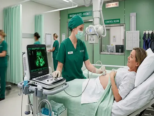
Bedside & Point-of-Care Ultrasound Imaging
The Portable Ultrasound Service at AS Medical Complex provides bedside ultrasound imaging for selected patients who require diagnostic assessment without unnecessary transportation to the main imaging department.
Portable ultrasound brings ultrasound technology directly to the patient’s location and can be particularly useful for patients who are:
-
Hospitalized
-
Acutely unwell
-
Less mobile
-
Recovering after surgery
-
Receiving emergency care
-
Requiring bedside reassessment
-
Difficult to transport safely
-
Requiring selected real-time clinical assessments
Point-of-care ultrasound – commonly referred to as POCUS – uses portable ultrasound technology at or near the patient’s bedside to provide real-time visualization of internal structures and help clinicians answer focused clinical questions.
Ultrasound uses high-frequency sound waves rather than X-rays to create images of structures inside the body.
It does not use ionizing radiation.
Depending on the clinical situation and available equipment, portable ultrasound may assist in assessment of:
-
Abdomen
-
Kidneys
-
Urinary bladder
-
Gallbladder
-
Selected liver abnormalities
-
Pelvic structures
-
Pleural fluid
-
Selected lung findings
-
Fluid within the abdomen
-
Selected vascular structures
-
Selected soft-tissue abnormalities
-
Other focused bedside clinical questions
Portable ultrasound should be used for an appropriate medical indication and performed or interpreted by healthcare professionals with suitable ultrasound training.
It should not automatically be considered a replacement for a complete Radiology Department ultrasound, CT Scan, MRI, X-Ray, echocardiography, or another specialized examination.
Our Portable Ultrasound Services
Depending on the patient’s condition, clinical indication, available equipment, and appropriately trained personnel, portable ultrasound services at AS Medical Complex may include:
-
Bedside ultrasound
-
Point-of-care ultrasound – POCUS
-
Portable abdominal ultrasound
-
Portable renal ultrasound
-
Portable urinary-bladder assessment
-
Portable hepatobiliary assessment
-
Selected gallbladder assessment
-
Selected liver assessment
-
Assessment for free abdominal fluid
-
Selected pelvic ultrasound
-
Selected urinary-retention assessment
-
Bladder-volume assessment where available
-
Selected pleural ultrasound
-
Assessment for pleural fluid
-
Selected lung ultrasound
-
Focused bedside assessment in Emergency care
-
Focused bedside assessment for hospitalized patients
-
Selected postoperative ultrasound assessment
-
Ultrasound assessment of selected superficial soft tissues
-
Selected vascular ultrasound assessment where capability is available
-
Doppler assessment where clinically appropriate and available
-
Ultrasound-guided procedure support where appropriate and specifically available
-
Follow-up bedside ultrasound
-
Referral for comprehensive departmental ultrasound when required
-
Referral for CT Scan, MRI, or other imaging where ultrasound is insufficient
The exact scope of portable ultrasound should be based on the equipment available at AS Medical Complex and the training and credentials of the clinician performing the examination.
What Is Portable Ultrasound?
Portable ultrasound uses a compact ultrasound system that can be moved to the patient rather than requiring the patient to travel to a fixed imaging room.
Depending on the system, portable equipment may include:
-
Cart-based ultrasound machines
-
Compact portable units
-
Laptop-style ultrasound systems
-
Handheld ultrasound devices
The basic imaging principle is the same as conventional ultrasound.
A transducer sends high-frequency sound waves into the body.
These sound waves interact with internal tissues and return to the transducer as echoes.
The ultrasound system processes the returning signals to create images in real time.
Portable Ultrasound vs Point-of-Care Ultrasound
The terms portable ultrasound and point-of-care ultrasound are related but are not always identical.
Portable Ultrasound
Portable ultrasound generally describes the mobility of the equipment.
The machine can be taken to:
-
Hospital bed
-
Emergency area
-
Procedure room
-
Other appropriate clinical location
Point-of-Care Ultrasound – POCUS
POCUS usually refers to a focused ultrasound examination performed by a trained healthcare professional at the point where the patient is receiving care.
Its purpose is often to answer a specific clinical question rapidly.
AIUM describes POCUS as bedside ultrasound that can support applications ranging from trauma assessment to procedure guidance.
A formal comprehensive ultrasound examination and a focused POCUS examination may therefore have different scopes.
Portable Ultrasound vs Comprehensive Radiology Ultrasound
A portable bedside examination may be appropriate when the immediate question is focused.
Examples include:
-
Is there significant fluid around the lung?
-
Is the bladder markedly distended?
-
Is there free fluid within the abdomen?
-
Is there an obvious gallbladder abnormality requiring further evaluation?
-
Is bedside reassessment useful in an unstable or immobile patient?
A comprehensive departmental ultrasound may provide:
-
More detailed organ assessment
-
More standardized imaging protocols
-
Additional transducers
-
More extensive Doppler imaging
-
Better positioning options
-
More complete documentation
-
Specialist radiology interpretation
Portable ultrasound should therefore complement rather than automatically replace comprehensive Radiology imaging.
Why Is Portable Ultrasound Useful?
Portable ultrasound can reduce the need to move selected patients from their clinical care area.
This can be useful when transportation is:
-
Difficult
-
Uncomfortable
-
Clinically undesirable
-
Logistically complicated
-
Potentially disruptive to monitoring or treatment
AIUM highlights portability as an important advantage of POCUS because it allows imaging to occur directly where the patient is receiving care.
Real-Time Imaging
Ultrasound produces images in real time.
This means the operator can observe:
-
Movement
-
Organ relationships
-
Fluid
-
Selected blood flow with Doppler
-
Changes during breathing
-
Selected dynamic abnormalities
This ability distinguishes ultrasound from static imaging such as a routine X-Ray.
No Ionizing Radiation
Portable ultrasound does not use ionizing radiation.
This is different from:
-
X-Ray
-
CT Scan
-
Fluoroscopy
Ultrasound instead uses sound-wave energy to generate images.
This can be particularly advantageous when repeated focused imaging is medically required.
However, ultrasound should still be used prudently rather than unnecessarily.
Ultrasound Safety
Diagnostic ultrasound has a long history of medical use and is generally considered safe when appropriately performed.
However, ultrasound introduces acoustic energy into the body.
FDA therefore recommends that ultrasound exposure be minimized while still obtaining the diagnostic information required, following the As Low As Reasonably Achievable – ALARA principle.
This is particularly important for:
-
Pregnancy
-
Doppler examinations
-
Repeated imaging
-
Prolonged examinations
The goal is not simply to scan for as long as possible.
The goal is to obtain the required medical information efficiently and safely.
Portable Abdominal Ultrasound
Portable abdominal ultrasound may help evaluate selected hospitalized or less-mobile patients with abdominal concerns.
Depending on the clinical question, bedside assessment may examine structures such as:
-
Liver
-
Gallbladder
-
Kidneys
-
Urinary bladder
-
Selected blood vessels
-
Abdominal fluid
-
Other accessible structures
Comprehensive abdominal ultrasound can provide images of organs and structures within the upper abdomen.
The quality and scope of a portable examination depend on:
-
Patient position
-
Body habitus
-
Bowel gas
-
Clinical condition
-
Available acoustic windows
-
Equipment
-
Operator experience
Abdominal Pain & Portable Ultrasound
Portable ultrasound may sometimes contribute to the assessment of abdominal pain.
Possible focused questions may involve:
-
Gallbladder abnormalities
-
Urinary obstruction
-
Bladder distension
-
Free abdominal fluid
-
Selected kidney abnormalities
-
Other focused concerns
However, abdominal pain has many possible causes.
Portable ultrasound cannot reliably exclude every condition.
Depending on the presentation, further investigation may require:
-
Comprehensive ultrasound
-
CT Scan
-
MRI
-
Laboratory tests
-
Endoscopy
-
Colonoscopy
-
Surgical assessment
The treating doctor determines the appropriate pathway.
Gallbladder Assessment
Ultrasound is commonly used to evaluate the gallbladder.
It may help identify selected abnormalities such as:
-
Gallstones
-
Gallbladder distension
-
Wall thickening
-
Fluid around the gallbladder
-
Other findings
RadiologyInfo lists gallstones and gallbladder inflammation among conditions commonly assessed with abdominal ultrasound.
Clinical diagnosis may still require:
-
Symptoms
-
Physical examination
-
Blood tests
-
Comprehensive imaging
-
Surgical consultation
Gallstones
Gallstones may be found incidentally in patients who have no symptoms.
Finding gallstones on ultrasound does not automatically mean that surgery is required.
Treatment depends on:
-
Symptoms
-
Complications
-
Gallbladder inflammation
-
Bile-duct obstruction
-
Pancreatitis
-
Other clinical factors
The imaging result should be reviewed by the treating clinician.
Suspected Acute Cholecystitis
Acute cholecystitis is inflammation of the gallbladder, frequently associated with gallstones.
Possible symptoms include:
-
Right upper abdominal pain
-
Fever
-
Nausea
-
Vomiting
-
Tenderness
Ultrasound may contribute important diagnostic information, but the diagnosis is based on the combination of clinical findings, laboratory results, and imaging.
Patients with severe symptoms require appropriate medical or surgical assessment.
Liver Assessment
Portable ultrasound may provide selected information regarding:
-
Liver size
-
Gross liver appearance
-
Selected focal abnormalities
-
Biliary dilation
-
Abdominal fluid
-
Other visible findings
However, comprehensive liver assessment may require:
-
Departmental ultrasound
-
Doppler
-
CT
-
MRI
-
Laboratory testing
-
Other investigations
depending on the suspected condition.
Fatty Liver
Ultrasound can sometimes show features suggesting fatty change within the liver.
However, ultrasound findings alone do not determine:
-
Exact severity of liver disease
-
Degree of fibrosis
-
Cause of liver abnormalities
Patients may require assessment involving:
-
Liver-function tests
-
Metabolic assessment
-
Gastroenterology
-
Dietitian support
-
Other investigations
Ascites
Ascites refers to abnormal fluid accumulation within the abdominal cavity.
Ultrasound can be highly sensitive for detecting free fluid and may help determine:
-
Presence of fluid
-
Distribution
-
Approximate extent
-
Suitable areas for selected procedures where appropriate
Possible causes of ascites include:
-
Liver disease
-
Heart disease
-
Cancer
-
Infection
-
Other medical conditions
Identifying fluid is only the beginning of the diagnostic process.
The underlying cause must be evaluated.
Portable Renal Ultrasound
Portable renal ultrasound may help evaluate selected abnormalities involving the kidneys.
Potential focused questions may include:
-
Is there urinary obstruction?
-
Is hydronephrosis present?
-
Are there gross structural abnormalities?
-
Is further comprehensive imaging required?
A renal ultrasound can examine the kidneys and urinary tract without ionizing radiation.
Hydronephrosis
Hydronephrosis means dilation of the kidney’s urine-collecting system, generally because urine flow is impaired.
Possible causes can include:
-
Urinary stones
-
Enlarged prostate
-
Ureteric obstruction
-
Pelvic masses
-
Other urinary conditions
Portable ultrasound may detect significant hydronephrosis, but further imaging may be required to determine the exact cause.
Kidney Stones
Some kidney stones can be identified using ultrasound, particularly when associated with urinary obstruction.
However, ultrasound does not detect every urinary stone equally well.
In selected adult patients with suspected acute kidney stones, non-contrast CT may provide more detailed assessment.
The appropriate investigation depends on:
-
Symptoms
-
Age
-
Pregnancy
-
Kidney function
-
Previous stone history
-
Clinical urgency
Urinary Bladder Ultrasound
Portable ultrasound may help evaluate the urinary bladder.
Potential indications include:
-
Suspected urinary retention
-
Bladder distension
-
Post-void residual assessment where appropriate
-
Selected urinary symptoms
-
Catheter-related clinical questions
The examination may help estimate whether a significant volume of urine remains within the bladder.
Urinary Retention
Urinary retention occurs when a patient cannot adequately empty the bladder.
Symptoms can include:
-
Difficulty passing urine
-
Lower abdominal discomfort
-
Reduced urine flow
-
Inability to urinate
-
Overflow leakage
Portable bladder ultrasound can sometimes provide rapid bedside information.
Severe acute retention may require:
-
Medical assessment
-
Catheterization
-
Urology evaluation
-
Investigation of the underlying cause
Urinary Catheter Assessment
Portable ultrasound may occasionally help when a patient with a urinary catheter has:
-
Reduced urine output
-
Lower abdominal distension
-
Concern about bladder filling
-
Other catheter-related problems
However, ultrasound does not replace clinical assessment of:
-
Catheter blockage
-
Kidney function
-
Hydration
-
Blood pressure
-
Other possible causes of reduced urine output
Portable Pelvic Ultrasound
Ultrasound of the pelvis can evaluate structures involving the urinary and reproductive systems.
RadiologyInfo describes pelvic ultrasound as a non-invasive, non-ionizing examination used to assess organs and structures within the lower abdomen and pelvis.
Depending on the patient and clinical indication, structures may include:
-
Urinary bladder
-
Uterus
-
Ovaries
-
Other pelvic structures
Portable transabdominal imaging may provide useful focused information, but detailed gynecological assessment may require a comprehensive departmental examination.
Female Pelvic Ultrasound
In women, pelvic ultrasound may be used to help evaluate selected:
-
Pelvic pain
-
Abnormal bleeding
-
Uterine abnormalities
-
Ovarian abnormalities
-
Pelvic masses
-
Other gynecological concerns
A complete pelvic assessment may use:
-
Transabdominal ultrasound
-
Transvaginal ultrasound where appropriate
Portable bedside equipment does not automatically mean every specialist gynecological ultrasound technique is available at the bedside.
Fibroids
Fibroids are benign muscular growths arising from the uterus.
Ultrasound can help assess:
-
Presence
-
Size
-
Location
-
Number of selected fibroids
However, complex fibroid disease may require:
-
Comprehensive pelvic ultrasound
-
MRI
-
Gynecology consultation
depending on symptoms and treatment planning.
Ovarian Abnormalities
Ultrasound can help identify selected ovarian abnormalities such as:
-
Cysts
-
Enlarged ovary
-
Masses
-
Other structural changes
An ovarian cyst does not automatically mean cancer.
Many ovarian cysts are benign.
Further management depends on:
-
Patient age
-
Menstrual status
-
Symptoms
-
Size
-
Ultrasound appearance
-
Other risk factors
Pelvic Pain
Pelvic pain can arise from many different organs and conditions.
Possible causes can involve:
-
Gynecological structures
-
Urinary tract
-
Gastrointestinal tract
-
Musculoskeletal system
Portable ultrasound may assist in selected circumstances but cannot exclude every serious cause.
Persistent, severe, or unexplained pelvic pain requires proper clinical evaluation.
Pregnancy & Portable Ultrasound
Ultrasound is widely used for medically indicated pregnancy imaging because it does not use ionizing radiation.
Portable ultrasound may sometimes be useful for a focused bedside obstetric assessment, particularly where immediate clinical information is required.
However, a focused bedside scan should not automatically be considered equivalent to a complete formal obstetric ultrasound.
A comprehensive obstetric study may require systematic assessment of:
-
Fetal anatomy
-
Growth
-
Placenta
-
Amniotic fluid
-
Gestational measurements
-
Other obstetric parameters
according to gestational stage and clinical indication.
Ultrasound in Early Pregnancy
Ultrasound may be used in selected early-pregnancy situations to assess:
-
Location of pregnancy
-
Gestational sac
-
Fetal cardiac activity where appropriate
-
Bleeding
-
Pain
-
Other obstetric concerns
The exact examination may require:
-
Transabdominal ultrasound
-
Transvaginal ultrasound
-
Serial hCG testing
-
Repeat ultrasound
depending on gestational age and clinical findings.
Ectopic Pregnancy
An ectopic pregnancy occurs when a pregnancy implants outside the normal uterine cavity.
It can become life-threatening if rupture causes internal bleeding.
Warning symptoms can include:
-
Significant pelvic or abdominal pain
-
Vaginal bleeding
-
Dizziness
-
Fainting
-
Shoulder-tip pain
-
Collapse
A patient with suspected ectopic pregnancy requires urgent Gynecology or Emergency assessment.
A portable ultrasound may contribute to initial evaluation, but it should not delay definitive specialist imaging or treatment.
Pregnancy Ultrasound Safety
Although diagnostic ultrasound is generally regarded as safe, professional guidance recommends using ultrasound only when medically indicated and minimizing acoustic exposure.
FDA recommends the ALARA principle, while AIUM specifically advises prudent use during pregnancy and discourages unnecessary nonmedical scanning.
Keepsake Pregnancy Scanning
Portable ultrasound should not be used solely to create entertainment or “keepsake” fetal images without a medical purpose.
AIUM strongly discourages nonmedical ultrasound use simply to view the fetus or obtain images outside a medically indicated examination.
At AS Medical Complex, pregnancy ultrasound should remain part of responsible medical care.
Doppler Ultrasound
Doppler ultrasound is a specialized ultrasound technique used to assess movement, especially blood flow.
It can help evaluate blood moving through:
-
Arteries
-
Veins
-
Selected organs
RadiologyInfo describes Doppler ultrasound as a technique that evaluates the movement of blood through vessels.
Availability of portable Doppler assessment depends on:
-
Device capability
-
Clinical indication
-
Operator expertise
Portable Doppler Assessment
Depending on available equipment and trained personnel, portable Doppler may assist selected focused questions involving:
-
Blood flow
-
Selected vessels
-
Organ perfusion
-
Other clinically appropriate assessments
However, a focused bedside Doppler study should not automatically replace a comprehensive vascular ultrasound examination.
Detailed vascular assessment may require:
-
Dedicated duplex ultrasound
-
Vascular laboratory equipment
-
Specialized sonographer or radiologist assessment
-
CT Angiography
-
MR Angiography
-
Other vascular investigations
Doppler During Pregnancy
Doppler imaging uses higher acoustic output than some basic ultrasound modes.
AIUM advises that pulsed Doppler should not be used routinely in early pregnancy without a medical indication and recommends minimizing exposure duration and acoustic output.
Pregnancy Doppler should therefore be used when there is a valid clinical reason.
Portable Lung Ultrasound
Portable ultrasound can be used in selected bedside assessment of the lungs and pleural spaces.
Focused lung ultrasound may help clinicians evaluate certain findings related to:
-
Pleural fluid
-
Pneumothorax in selected settings
-
Interstitial fluid patterns
-
Selected lung consolidation
-
Other focused respiratory questions
AIUM recognizes lung ultrasound as an important point-of-care application in appropriate clinical settings.
Lung ultrasound does not replace:
-
Chest X-Ray
-
CT Chest
-
Pulmonary Function Testing
-
Other investigations
when those tests are clinically required.
Pleural Effusion
A pleural effusion is fluid accumulating in the space surrounding the lung.
Ultrasound can help:
-
Detect pleural fluid
-
Estimate its location
-
Assess its accessibility
-
Guide selected procedures where properly available
Causes can include:
-
Heart failure
-
Infection
-
Liver disease
-
Cancer
-
Other conditions
Finding pleural fluid does not establish the underlying diagnosis.
Pneumothorax
A pneumothorax occurs when air accumulates between the lung and chest wall.
Focused lung ultrasound can support diagnosis in appropriately trained hands and selected clinical settings.
However, suspected pneumothorax may also require:
-
Chest X-Ray
-
CT
-
Emergency treatment
depending on the patient’s stability.
A patient with severe breathing difficulty or chest trauma requires urgent medical assessment.
Portable Ultrasound in Emergency Care
Point-of-care ultrasound can be useful in Emergency settings because imaging can be performed rapidly at the bedside.
AIUM identifies applications including trauma assessment and procedure guidance.
Depending on the emergency and trained personnel available, bedside ultrasound may help address selected questions involving:
-
Internal free fluid
-
Cardiac activity within appropriately trained scope
-
Pleural fluid
-
Pneumothorax
-
Bladder distension
-
Abdominal aortic assessment
-
Other urgent focused questions
A bedside ultrasound should support—not replace—the wider Emergency assessment.
FAST Examination
In trauma care, a focused ultrasound examination may be used to look for free fluid in selected body spaces.
This type of examination is commonly referred to as FAST – Focused Assessment with Sonography in Trauma.
Where an extended protocol is used, lung assessment may also be incorporated.
A FAST examination is a focused emergency examination.
It does not exclude every abdominal, chest, or pelvic injury.
A trauma patient may still require CT even when bedside ultrasound is negative.
FAST/eFAST should only be advertised as an AS Medical Complex service if appropriately trained clinicians and protocols are available.
Major Trauma
Patients with significant trauma may require:
-
Emergency stabilization
-
X-Ray
-
CT Scan
-
Laboratory tests
-
Surgical consultation
-
Other investigations
Portable ultrasound may provide rapid information while these other assessments are arranged.
It should not delay definitive imaging when CT is clinically necessary.
Portable Ultrasound for Inpatients
Hospitalized patients may benefit from bedside ultrasound when moving them to the Radiology Department is difficult.
Possible clinical uses may include selected evaluation of:
-
Abdominal fluid
-
Urinary retention
-
Kidney obstruction
-
Pleural fluid
-
Selected respiratory findings
-
Other focused clinical problems
The examination should be ordered for a clear clinical reason.
Portable Ultrasound After Surgery
Selected postoperative patients may undergo bedside ultrasound when there is concern about:
-
Fluid accumulation
-
Urinary retention
-
Pleural fluid
-
Selected abdominal abnormalities
-
Other postoperative issues
However, suspected major postoperative complications can require:
-
CT Scan
-
Laboratory testing
-
Surgical assessment
-
Other imaging
Portable ultrasound should not delay further investigation if the patient’s condition is deteriorating.
Portable Ultrasound & Dialysis
Patients receiving dialysis may occasionally require ultrasound assessment because of:
-
Reduced urine output
-
Abdominal symptoms
-
Fluid-related questions
-
Other medical problems
Specialized evaluation of an AV fistula or graft generally requires appropriate vascular or Doppler ultrasound expertise and should not be assumed to be part of a basic portable ultrasound service.
Portable Ultrasound & Kidney Failure
Ultrasound may help identify gross structural findings such as:
-
Hydronephrosis
-
Kidney size
-
Bladder distension
-
Selected urinary abnormalities
However, ultrasound cannot determine kidney function by itself.
Kidney function assessment may also require:
-
Creatinine
-
eGFR
-
Electrolytes
-
Urine testing
-
Nephrology consultation
Portable Ultrasound for Soft-Tissue Problems
Portable ultrasound may help evaluate selected superficial soft-tissue problems.
Examples may include:
-
Fluid collections
-
Selected abscesses
-
Cysts
-
Other superficial abnormalities
Ultrasound can provide good visualization of many soft tissues without ionizing radiation.
Some abnormalities may still require:
-
Formal ultrasound
-
MRI
-
CT
-
Surgical assessment
-
Biopsy
Abscess Assessment
An abscess is a collection of infected fluid.
Ultrasound can help determine whether a superficial swelling appears:
-
Predominantly fluid-filled
-
Solid
-
Mixed
Clinical signs may include:
-
Pain
-
Redness
-
Warmth
-
Swelling
-
Fever
An abscess may require drainage and other treatment.
Imaging alone does not treat the infection.
Ultrasound-Guided Procedures
Ultrasound can help visualize anatomy in real time and may be used to guide selected procedures. RadiologyInfo recognizes ultrasound guidance as an established application for procedures such as biopsies and other interventions.
Depending on clinical service availability, possible uses may include guidance for selected:
-
Vascular-access procedures
-
Fluid aspiration
-
Drainage
-
Other interventions
Such procedures require appropriate:
-
Clinical expertise
-
Sterile technique
-
Consent
-
Monitoring
-
Equipment
They should only be advertised as AS Medical Complex services after onsite capability is confirmed.
Portable Ultrasound for Vascular Access
Point-of-care ultrasound can assist trained clinicians in selected vascular-access procedures by helping visualize:
-
Veins
-
Arteries
-
Surrounding structures
AIUM specifically recognizes vascular-access guidance among POCUS applications.
This may be useful in patients with difficult vascular access.
Portable Ultrasound Limitations
Portable ultrasound has important limitations.
Image quality can be affected by:
-
Obesity
-
Bowel gas
-
Wounds
-
Dressings
-
Patient positioning
-
Inability to cooperate
-
Pain
-
Limited acoustic windows
-
Equipment limitations
-
Operator experience
Ultrasound waves do not pass effectively through air-filled lung or dense bone, which limits assessment of some structures. Comprehensive or alternative imaging may therefore be required.
Operator Dependence
Ultrasound is more operator-dependent than many imaging techniques.
The diagnostic value of the examination depends partly on:
-
Selecting the correct transducer
-
Proper probe positioning
-
Understanding anatomy
-
Obtaining adequate views
-
Recognizing artifacts
-
Interpreting findings appropriately
AIUM maintains specific training, practice-parameter, documentation, and quality requirements for point-of-care ultrasound.
Portable equipment alone does not make an ultrasound examination reliable.
Portable Ultrasound Does Not Replace CT
CT may be preferred when doctors need detailed assessment of:
-
Major trauma
-
Internal organs
-
Complex abdominal disease
-
Acute intracranial disease
-
Lung abnormalities
-
Complex fractures
-
Other deep structures
CT is also less affected by many of the acoustic limitations that affect ultrasound.
A patient should therefore proceed to CT when medically indicated even if a bedside ultrasound has already been performed.
Portable Ultrasound Does Not Replace MRI
MRI may provide more detailed evaluation of:
-
Brain
-
Spinal cord
-
Nerves
-
Intervertebral discs
-
Ligaments
-
Tendons
-
Muscles
-
Complex pelvic disease
-
Other selected soft tissues
Portable ultrasound and MRI answer different clinical questions.
Portable Ultrasound Does Not Replace Echocardiography
Focused bedside cardiac ultrasound can sometimes help trained clinicians answer limited immediate questions.
However, it should not automatically be regarded as equivalent to a formal echocardiogram.
Formal echocardiography may provide detailed assessment of:
-
Heart chambers
-
Ventricular function
-
Heart valves
-
Doppler measurements
-
Pulmonary pressures
-
Other cardiac parameters
Patients who require comprehensive cardiac assessment may need the dedicated Echo & ETT Service or Cardiology assessment.
Portable Ultrasound Does Not Replace Formal Obstetric Ultrasound
A focused bedside pregnancy scan may answer a limited clinical question.
A formal obstetric ultrasound may require systematic evaluation of:
-
Pregnancy dating
-
Fetal anatomy
-
Growth
-
Placenta
-
Amniotic fluid
-
Other pregnancy-related structures
Portable ultrasound should therefore not be promoted as a substitute for comprehensive antenatal imaging.
Preparation for Portable Ultrasound
Preparation depends on the area being examined.
Some portable examinations may require little or no preparation.
Others may benefit from:
-
Fasting
-
A full bladder
-
Specific positioning
-
Other instructions
Whenever possible, patients should follow the preparation instructions provided by the healthcare team.
Preparation for Abdominal Ultrasound
For some abdominal ultrasound examinations, fasting may improve visualization by reducing bowel gas and helping assessment of the gallbladder.
Patients may be asked not to eat for a specified period before the study.
The exact requirement depends on the clinical question.
In urgent bedside situations, imaging may still be performed without ideal preparation if medically necessary.
Preparation for Pelvic Ultrasound
A transabdominal pelvic ultrasound often provides better visualization when the urinary bladder is appropriately filled.
RadiologyInfo notes that patients may be instructed to drink water before some pelvic ultrasound examinations.
However, bedside examinations may need to proceed according to the immediate clinical situation.
What Happens During Portable Ultrasound?
During a bedside ultrasound:
-
Patient identity is confirmed.
-
The clinical question is reviewed.
-
The patient is positioned as safely as possible.
-
The appropriate area is exposed.
-
Ultrasound gel is applied to the skin.
-
A transducer is moved over the area being examined.
-
Real-time images are obtained.
-
Selected measurements may be taken.
-
Images may be stored according to the examination and hospital protocol.
The examination is generally non-invasive and usually painless, although pressure from the probe may be uncomfortable over a tender area.
Ultrasound Gel
A water-based gel is commonly placed between the transducer and skin.
The gel helps eliminate air between the probe and skin so that ultrasound waves can pass effectively into the body.
It is removed after the examination.
How Long Does Portable Ultrasound Take?
The duration varies according to:
-
Clinical question
-
Number of structures being examined
-
Patient condition
-
Image quality
-
Need for Doppler
-
Difficulty obtaining appropriate views
A focused point-of-care examination may be relatively brief.
A comprehensive examination generally requires more time.
AS Medical Complex should therefore avoid advertising one fixed examination time for every portable ultrasound.
Portable Ultrasound Results
How results are communicated depends on the type of examination.
For a focused POCUS examination, findings may be immediately incorporated into the treating clinician’s assessment.
For a formal portable Radiology examination, images may require review and reporting by an appropriately qualified imaging specialist.
AIUM/ACR practice guidance emphasizes appropriate documentation and retention of ultrasound examination information.
Focused Result vs Formal Radiology Report
Patients should understand the distinction between:
Focused Bedside Assessment
Designed to answer a limited clinical question.
Comprehensive Ultrasound Examination
Designed to systematically evaluate an organ or anatomical region.
A focused bedside result does not always constitute a complete evaluation of all surrounding structures.
Additional formal imaging may therefore still be recommended.
Normal Portable Ultrasound Does Not Exclude Every Disease
A normal bedside ultrasound does not guarantee that no medical condition is present.
Ultrasound can be limited by:
-
Patient anatomy
-
Bowel gas
-
Depth
-
Body habitus
-
Disease location
-
Equipment
-
Examination scope
A negative focused examination may therefore need to be followed by:
-
Comprehensive ultrasound
-
CT
-
MRI
-
Laboratory tests
-
Endoscopy
-
Other investigations
when clinical concern remains.
Incidental Findings
Portable ultrasound may sometimes reveal an unexpected abnormality unrelated to the original reason for imaging.
Depending on the finding, further assessment may involve:
-
Formal Radiology ultrasound
-
CT Scan
-
MRI
-
Laboratory testing
-
Specialist consultation
An incidental finding does not automatically mean serious disease.
Infection Prevention & Ultrasound Equipment
Ultrasound transducers make direct contact with patients and therefore require appropriate cleaning and disinfection procedures.
AIUM quality standards specifically require ultrasound practices to maintain procedures for preventing infection transmission and cleaning/disinfecting ultrasound equipment and transducers between patients.
Portable equipment should follow the same infection-control principles even though it moves between clinical areas.
Bedside Ultrasound & Isolation Patients
Portable ultrasound may be particularly useful in selected isolation settings because the equipment can be brought to the patient.
However, infection-control precautions are essential.
These may include:
-
Appropriate protective equipment
-
Correct probe cleaning
-
Disinfection
-
Equipment cleaning
-
Safe handling between patients
The exact precautions depend on the infection and hospital policy.
Quality Assurance
Portable ultrasound systems require appropriate quality assurance.
FDA recommends regular quality-control testing so that ultrasound equipment continues to function properly.
Quality care also involves:
-
Trained operators
-
Proper equipment maintenance
-
Appropriate documentation
-
Correct transducer selection
-
Infection prevention
-
Clinical correlation
Portable Ultrasound & Radiology
The Radiology Department may coordinate portable ultrasound when a formal bedside imaging examination is required.
Radiology involvement may support:
-
Appropriate protocol selection
-
Image acquisition
-
Interpretation
-
Reporting
-
Recommendation for additional imaging
Some focused POCUS examinations may instead be performed directly by appropriately trained treating clinicians.
Portable Ultrasound & Emergency Services
Emergency clinicians may use portable ultrasound to help rapidly evaluate selected urgent clinical questions.
Depending on available expertise, bedside ultrasound may complement:
-
Physical examination
-
Laboratory tests
-
ECG
-
X-Ray
-
CT
-
Specialist consultation
It should be considered part of a broader Emergency assessment.
Portable Ultrasound & General Medicine
General Medicine may request bedside ultrasound for selected hospitalized patients with concerns such as:
-
Abdominal symptoms
-
Urinary retention
-
Fluid accumulation
-
Selected respiratory problems
-
Other focused conditions
The result may determine whether further comprehensive imaging is required.
Portable Ultrasound & Gastroenterology
Gastroenterology-related bedside ultrasound may help evaluate selected questions involving:
-
Liver
-
Gallbladder
-
Ascites
-
Other abdominal abnormalities
More complex gastrointestinal disease may require:
-
Full abdominal ultrasound
-
CT
-
MRI
-
MRCP
-
Endoscopy
-
Colonoscopy
depending on the problem.
Portable Ultrasound & Nephrology
Nephrology may use ultrasound information in patients with:
-
Acute Kidney Injury
-
Chronic Kidney Disease
-
Suspected urinary obstruction
-
Reduced urine output
-
Bladder distension
-
Other kidney-related concerns
Kidney function itself must still be assessed through appropriate laboratory and clinical evaluation.
Portable Ultrasound & Urology
Urology may use ultrasound in selected cases involving:
-
Urinary retention
-
Hydronephrosis
-
Bladder abnormalities
-
Kidney abnormalities
-
Other urinary conditions
Further investigation may require:
-
Formal renal ultrasound
-
CT
-
MRI
-
Endoscopy of the urinary tract
-
Other specialist testing
Portable Ultrasound & Gynecology
Gynecology may use ultrasound to assess selected:
-
Pelvic pain
-
Pregnancy-related concerns
-
Uterine abnormalities
-
Ovarian abnormalities
A comprehensive Gynecology ultrasound may require specialized transabdominal or transvaginal imaging rather than a limited portable examination.
Portable Ultrasound & General Surgery
General Surgery may use bedside ultrasound information in selected patients with:
-
Gallbladder symptoms
-
Abdominal fluid
-
Trauma
-
Postoperative concerns
-
Other surgical problems
If acute surgical disease is suspected, ultrasound findings should be considered alongside:
-
Examination
-
Laboratory testing
-
CT
-
Other imaging
Portable Ultrasound & Pulmonology
Pulmonology may use ultrasound for selected bedside assessment of:
-
Pleural fluid
-
Selected lung-surface abnormalities
-
Other focused questions
Deeper lung disease may require:
-
Chest X-Ray
-
CT Chest
-
HRCT
-
Pulmonary Function Testing
-
Other investigations
Portable Ultrasound & Cardiology
Focused cardiac ultrasound may sometimes provide limited bedside information when performed by appropriately trained clinicians.
However, patients requiring detailed heart assessment may need:
-
ECG
-
Echocardiography
-
ETT
-
Holter monitoring
-
Other Cardiology investigations
depending on symptoms.
Portable Ultrasound & Pediatrics
Portable ultrasound may be particularly useful in selected children because it does not use ionizing radiation.
RadiologyInfo notes that ultrasound can be useful for evaluating abdominal, pelvic, and scrotal pain in children.
However, pediatric imaging should still be selected according to:
-
Age
-
Symptoms
-
Clinical question
-
Need for comprehensive examination
When Should Portable Ultrasound Be Considered?
Portable ultrasound may be useful when:
-
A hospitalized patient needs focused ultrasound assessment.
-
Transporting the patient to Radiology is difficult.
-
A patient requires rapid bedside evaluation.
-
Urinary retention is suspected.
-
Significant bladder distension is suspected.
-
Free abdominal fluid is being assessed.
-
Pleural fluid is suspected.
-
A selected abdominal or renal question can be assessed at the bedside.
-
Follow-up bedside assessment is clinically useful.
-
The treating clinician needs real-time ultrasound information to guide further investigation.
The clinical team should determine whether a portable examination is sufficient or whether the patient needs formal Radiology imaging.
When Is Comprehensive Ultrasound More Appropriate?
A formal departmental ultrasound may be preferable when:
-
Detailed organ assessment is required.
-
Extensive measurements are needed.
-
Specialized Doppler evaluation is required.
-
A full obstetric examination is needed.
-
Detailed vascular assessment is required.
-
A focused portable study is inconclusive.
-
Image quality is inadequate at the bedside.
-
Specialist radiology interpretation is required.
Portable ultrasound should not be used simply because moving the patient is inconvenient when a comprehensive examination is medically necessary.
When Is CT or MRI More Appropriate?
Further imaging may be required when ultrasound cannot adequately evaluate:
-
Deep abdominal structures
-
Major trauma
-
Complex tumors
-
Brain
-
Spinal cord
-
Many lung abnormalities
-
Complex postoperative complications
-
Detailed vascular anatomy
-
Other structures limited by ultrasound
The treating clinician and Radiology team should choose the most appropriate imaging modality.
When Is Emergency Care Required?
Portable ultrasound should not delay emergency treatment when a patient has:
-
Severe breathing difficulty
-
Severe chest pain
-
Major trauma
-
Loss of consciousness
-
Severe internal or external bleeding
-
Shock
-
Sudden neurological weakness
-
Severe abdominal pain with collapse
-
Suspected ruptured ectopic pregnancy
-
Severe infection with deterioration
-
Other potentially life-threatening symptoms
These patients require immediate clinical stabilization.
Portable ultrasound may form part of the Emergency assessment, but additional treatment and imaging may be required.
Multidisciplinary Portable Ultrasound Care
Depending on the clinical question and findings, portable ultrasound care may involve:
-
Radiology
-
Emergency Services
-
General Medicine
-
Gastroenterology
-
Nephrology
-
Urology
-
Gynecology
-
General & Laparoscopic Surgery
-
Pulmonology
-
Cardiology
-
Pediatrics
-
Critical-care teams where applicable
-
Other appropriate specialties
Portable ultrasound is therefore best viewed as an extension of patient care rather than an isolated imaging test.
Follow-Up After Portable Ultrasound
The next step depends on:
-
Clinical condition
-
Ultrasound findings
-
Quality of the examination
-
Scope of the bedside study
Possible next steps may include:
-
No additional imaging
-
Medical treatment
-
Formal Radiology ultrasound
-
Doppler ultrasound
-
CT Scan
-
MRI
-
X-Ray
-
Laboratory investigations
-
Specialist consultation
-
Procedure
-
Surgery
-
Repeat ultrasound
Significant findings should be reviewed by the healthcare professional responsible for the patient.
Repeat Portable Ultrasound
One advantage of ultrasound is that focused examinations can sometimes be repeated when clinically useful without exposing the patient to ionizing radiation.
However, repeated scanning should still have a medical purpose.
FDA and AIUM recommend prudent ultrasound use and minimizing unnecessary acoustic exposure while obtaining the required diagnostic information.
Portable Ultrasound FAQs
What is portable ultrasound?
Portable ultrasound uses compact ultrasound equipment that can be brought directly to the patient’s bedside. It creates real-time images using sound waves and does not use ionizing radiation. Point-of-care ultrasound may be particularly useful when a trained clinician needs to answer a focused clinical question without unnecessarily moving the patient.
Is portable ultrasound the same as a complete ultrasound in the Radiology Department?
Not always. Portable ultrasound can be used for a formal bedside study or a focused point-of-care examination. A focused POCUS examination is often designed to answer a specific clinical question, whereas a comprehensive Radiology ultrasound may systematically evaluate an entire organ or anatomical region. Additional formal imaging may therefore still be required.
Is portable ultrasound safe?
Diagnostic ultrasound is generally considered safe when appropriately used and does not expose the patient to ionizing radiation. However, ultrasound introduces acoustic energy into tissues, so FDA recommends using the lowest exposure necessary to obtain adequate diagnostic information according to the ALARA principle.
Can portable ultrasound be used during pregnancy?
Ultrasound is commonly used for medically indicated pregnancy imaging because it does not use ionizing radiation. A focused bedside scan may sometimes provide useful immediate information, but it does not automatically replace a complete formal obstetric ultrasound. AIUM recommends prudent medically indicated use and discourages unnecessary nonmedical fetal scanning.
Can portable ultrasound detect kidney stones or gallstones?
Ultrasound can identify many gallstones and may detect some kidney stones or indirect evidence such as urinary obstruction. However, it does not identify every urinary stone, and CT may be required in selected patients. The appropriate imaging method depends on symptoms, clinical findings, pregnancy status, and other factors. Abdominal ultrasound is commonly used to assess gallstones and selected kidney abnormalities.
Does a normal portable ultrasound mean that nothing is wrong?
No. A focused portable ultrasound has a defined and sometimes limited scope. Image quality may also be affected by bowel gas, body habitus, patient position, and the location of disease. If clinical concern remains, the doctor may recommend comprehensive ultrasound, CT Scan, MRI, laboratory testing, or another investigation.
Who should perform and interpret portable ultrasound?
Portable ultrasound should be performed and interpreted by healthcare professionals with appropriate education, training, and competency for the type of examination being performed. AIUM maintains specific training, practice, documentation, and quality standards for point-of-care ultrasound.
Portable Ultrasound Services at AS Medical Complex
The Portable Ultrasound Service at AS Medical Complex is designed to bring appropriate ultrasound imaging closer to patients who may benefit from bedside assessment.
From focused abdominal, kidney, urinary-bladder, pelvic, pleural, lung, and selected bedside assessments to point-of-care ultrasound for hospitalized and Emergency patients, our goal is to provide timely imaging information while minimizing unnecessary patient movement.
Portable ultrasound uses sound waves and does not involve ionizing radiation, but safe and effective use still depends on appropriate clinical indications, trained operators, suitable equipment, correct documentation, infection prevention, and responsible control of ultrasound exposure.
A portable ultrasound examination should also be understood within its limitations. Some patients will require a comprehensive Radiology ultrasound, Doppler study, CT Scan, MRI, X-Ray, laboratory testing, or specialist assessment to establish the diagnosis.
Portable ultrasound findings can be coordinated with the relevant services at AS Medical Complex, including Radiology, Emergency Services, General Medicine, Gastroenterology, Nephrology, Urology, Gynecology, General & Laparoscopic Surgery, Pulmonology, Cardiology, Pediatrics, and other specialties according to clinical need.
Contact the Portable Ultrasound Service at AS Medical Complex for clinically indicated bedside ultrasound assessment for hospitalized, less-mobile, Emergency, postoperative, and other appropriately selected patients.