Breast Surgery at AS Medical Complex
Contact hospital
- Availability
Contact hospital
- Booking
WhatsApp / call
- Location
Islamabad
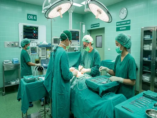
Comprehensive Breast Disease, Diagnostic & Surgical Care
The Breast Surgery Department at AS Medical Complex provides comprehensive assessment, diagnosis, surgical consultation, treatment planning, operative care, and follow-up for patients with benign and malignant breast conditions.
Breast symptoms are common and do not automatically indicate cancer. Breast lumps, breast pain, nipple discharge, infections, cysts, fibroadenomas, hormonal changes, and other benign conditions can produce symptoms requiring evaluation.
At the same time, persistent or suspicious breast changes require appropriate investigation so that breast cancer or another important condition can be identified as early as possible.
Our approach focuses on accurate diagnosis, appropriate imaging, biopsy when clinically indicated, careful surgical planning, preservation of healthy breast tissue where appropriate, multidisciplinary cancer care, and structured follow-up.
Our Breast Surgery Services
The Breast Surgery Department at AS Medical Complex may provide assessment and management for:
-
Breast lumps
-
Newly detected breast masses
-
Benign breast disease
-
Fibroadenoma
-
Breast cysts
-
Fibrocystic breast changes
-
Breast pain
-
Nipple discharge
-
Bloody nipple discharge
-
Nipple inversion
-
Breast skin changes
-
Breast infections
-
Mastitis
-
Breast abscess
-
Recurrent breast abscess
-
Abnormal mammography findings
-
Abnormal breast ultrasound findings
-
Abnormal breast MRI findings
-
Breast biopsy assessment
-
Core needle biopsy coordination
-
Image-guided biopsy coordination
-
Excisional breast biopsy
-
High-risk breast lesions
-
Ductal abnormalities
-
Breast cancer assessment
-
Ductal Carcinoma In Situ – DCIS
-
Invasive breast cancer
-
Breast-conserving surgery
-
Lumpectomy
-
Wide local excision
-
Mastectomy
-
Sentinel lymph-node biopsy
-
Axillary lymph-node surgery where clinically indicated
-
Male breast lumps
-
Gynecomastia assessment
-
Male breast cancer
-
Breast reconstruction coordination
-
Post-mastectomy care
-
Postoperative wound care
-
Breast surgical second opinions
-
Long-term follow-up after breast surgery
The exact investigation or surgical procedure recommended depends on the patient’s diagnosis, imaging, pathology, disease stage, general health, and individual treatment requirements.
Breast Lumps
A breast lump is one of the most common reasons patients seek breast assessment.
Breast lumps can develop because of several conditions, including:
-
Fibroadenoma
-
Breast cyst
-
Hormonal changes
-
Infection
-
Fat necrosis
-
Benign tissue changes
-
Breast cancer
-
Other breast conditions
A new breast lump should be properly evaluated rather than diagnosed by touch alone.
Assessment generally considers:
-
Patient age
-
Duration of the lump
-
Change in size
-
Pain or tenderness
-
Menstrual relationship
-
Pregnancy or breastfeeding
-
Family history
-
Previous breast conditions
-
Clinical examination
-
Imaging findings
Further investigation may be required when a lump has suspicious or uncertain features.
Triple Assessment of a Breast Lump
Breast abnormalities are often evaluated using a combination of:
-
Clinical examination
-
Breast imaging
-
Tissue diagnosis when required
Depending on the patient, imaging may involve:
-
Breast ultrasound
-
Mammography
-
MRI in selected situations
If imaging or examination identifies an abnormal area requiring tissue diagnosis, a biopsy may be recommended.
Image-guided breast biopsy can use ultrasound, mammography, or MRI to accurately target an abnormal area.
Breast Examination
A clinical breast assessment may include examination of:
-
Both breasts
-
Nipples
-
Breast skin
-
Armpits
-
Lymph-node areas around the breast
-
Areas above and below the collarbone when appropriate
The surgeon may assess:
-
Lump size
-
Lump mobility
-
Skin changes
-
Nipple changes
-
Tenderness
-
Swelling
-
Lymph nodes
-
Previous surgical scars
Clinical examination alone cannot reliably determine the cause of every breast lump, so imaging or biopsy may sometimes be required.
Mammography
A mammogram uses low-dose X-rays to produce detailed breast images.
Mammography can help identify abnormalities including:
-
Breast masses
-
Calcifications
-
Architectural changes
-
Other suspicious breast findings
Mammography is an important primary breast-cancer screening tool and can detect some abnormalities before they are large enough to be felt during examination.
The appropriate use of mammography depends on:
-
Age
-
Symptoms
-
Previous imaging
-
Breast density
-
Personal risk
-
Family history
-
Other clinical factors
Diagnostic Mammography
A diagnostic mammogram may be recommended when a patient has:
-
A breast lump
-
Nipple discharge
-
Skin changes
-
An abnormal screening mammogram
-
A previously identified breast abnormality
-
Other concerning symptoms
Additional views may be performed to examine a particular area more closely.
Breast Ultrasound
Breast ultrasound uses sound waves to produce images of breast tissue.
It can be particularly useful for:
-
Evaluating breast lumps
-
Distinguishing fluid-filled cysts from solid lesions
-
Examining younger patients
-
Evaluating areas of pain
-
Guiding needle biopsy
-
Assessing selected abnormalities found on mammography
Ultrasound and mammography often provide complementary information rather than replacing one another.
Breast MRI
Breast MRI may be used in selected patients when more detailed imaging is clinically required.
Possible indications may include:
-
Selected high-risk patients
-
Further evaluation of known breast cancer
-
Assessment of certain difficult imaging findings
-
Evaluation of breast implants in specific situations
-
Other specialist indications
MRI is not required for every breast lump or every patient diagnosed with breast cancer.
Breast Density
Breast tissue varies in density.
Dense breast tissue can make some abnormalities more difficult to identify on mammography and is also associated with increased breast-cancer risk.
Patients with dense breasts should discuss their individual imaging and risk profile with their treating clinician rather than assuming that one additional test is automatically necessary for everyone.
Breast Biopsy
A breast biopsy removes cells or tissue from an abnormal area so that the tissue can be examined by a pathologist.
Biopsy may be recommended when:
-
A lump appears suspicious
-
Imaging shows an abnormal area
-
A lesion has changed
-
The diagnosis remains uncertain
-
Cancer needs to be excluded or confirmed
Biopsy does not automatically mean that cancer is present.
Its purpose is to establish an accurate diagnosis.
Core Needle Biopsy
A core needle biopsy removes small samples of tissue using a hollow needle.
It may be performed using imaging guidance such as:
-
Ultrasound
-
Mammography
-
MRI
NCI identifies image-guided core needle biopsy as an important method for obtaining tissue from breast abnormalities, including lesions that cannot easily be felt.
Pathology results help guide further management.
Fine Needle Aspiration
Fine Needle Aspiration may be used in selected breast conditions to obtain cells or fluid using a thin needle.
It may sometimes be useful for:
-
Selected cysts
-
Certain lymph nodes
-
Other appropriate breast abnormalities
The choice between fine needle aspiration, core biopsy, and surgical biopsy depends on the clinical situation.
Excisional Breast Biopsy
An excisional biopsy involves surgically removing all or most of a breast lesion for pathological examination.
It may be considered when:
-
Needle biopsy does not provide adequate diagnosis
-
Imaging and pathology findings do not match
-
A particular lesion requires complete removal
-
The abnormality is symptomatic
-
There is another appropriate surgical indication
Many breast abnormalities can now be diagnosed with needle biopsy, so open surgical biopsy is not automatically necessary for every lump.
Benign Breast Disease
Not all breast diseases are cancerous.
Benign breast conditions may include:
-
Fibroadenoma
-
Breast cysts
-
Fibrocystic changes
-
Breast infection
-
Fat necrosis
-
Intraductal papilloma
-
Some inflammatory conditions
-
Other non-cancerous lesions
Management depends on symptoms, imaging, biopsy results, age, lesion size, growth, and individual risk factors.
The American Society of Breast Surgeons maintains specific clinical guidance for benign breast lesions because different benign abnormalities require different approaches rather than routine surgical removal of every lesion.
Fibroadenoma
A fibroadenoma is a common benign solid breast lesion, particularly in younger women.
It may feel:
-
Firm
-
Smooth
-
Mobile
-
Well-defined
Some fibroadenomas remain stable or become smaller with time.
Others may enlarge.
Assessment may involve:
-
Clinical examination
-
Ultrasound
-
Mammography depending on age
-
Core biopsy when appropriate
Not every fibroadenoma requires surgery.
When May Fibroadenoma Surgery Be Considered?
Surgical removal may be considered in selected patients when:
-
The diagnosis remains uncertain
-
The lesion is growing
-
It becomes large
-
It causes significant symptoms
-
Imaging or pathology has concerning features
-
The patient and surgeon determine that removal is appropriate
Current breast-surgery guidance generally discourages routine surgical excision of every small biopsy-proven fibroadenoma without another indication.
Breast Cysts
Breast cysts are fluid-filled structures.
Some patients have:
-
One cyst
-
Multiple cysts
-
Recurrent cysts
Symptoms may include:
-
A palpable lump
-
Tenderness
-
Fluctuation with the menstrual cycle
-
Breast discomfort
Ultrasound is often helpful in determining whether a lump is cystic or solid.
Some simple cysts require no treatment.
Selected painful or concerning cysts may require aspiration or further investigation.
Fibrocystic Breast Changes
Some patients experience breast lumpiness, tenderness, or discomfort related to benign changes in breast tissue.
Symptoms may fluctuate with hormonal cycles.
Assessment is important when there is:
-
A new dominant lump
-
Persistent focal pain
-
Nipple discharge
-
Skin change
-
An abnormal imaging finding
New symptoms should not simply be attributed to hormonal changes without appropriate evaluation.
Breast Pain
Breast pain is common and can occur for many reasons.
Pain may be:
-
Cyclical
-
Non-cyclical
-
Localized
-
Diffuse
-
Related to chest-wall structures rather than the breast itself
Possible causes include:
-
Hormonal changes
-
Breast cysts
-
Infection
-
Musculoskeletal pain
-
Trauma
-
Medication effects
-
Other breast conditions
Breast cancer is not the most common cause of breast pain, but persistent focal pain or pain associated with another abnormality should be assessed.
Nipple Discharge
Nipple discharge can have benign or more concerning causes.
Assessment may be particularly important when discharge is:
-
Bloody
-
Clear and spontaneous
-
From one breast
-
From a single duct
-
Associated with a breast lump
-
Associated with nipple or skin changes
The surgeon may recommend:
-
Clinical examination
-
Ultrasound
-
Mammography
-
MRI in selected cases
-
Biopsy or other investigation
depending on the findings.
Nipple Inversion
Some people naturally have inverted nipples.
However, new nipple inversion or a noticeable change in nipple position should be assessed, especially when associated with:
-
A lump
-
Skin changes
-
Discharge
-
Breast distortion
A change does not automatically indicate cancer but may require imaging or further investigation.
Breast Skin Changes
Breast skin changes requiring assessment may include:
-
Persistent redness
-
Thickening
-
Dimpling
-
Puckering
-
Ulceration
-
Persistent rash around the nipple
-
Orange-peel-like texture
-
Rapid breast swelling
The FDA advises medical evaluation for changes such as a breast lump, nipple leakage, or changes in the appearance of the breast skin or nipple.
Mastitis
Mastitis refers to inflammation of breast tissue and frequently occurs during breastfeeding, although it can occur in other circumstances.
Symptoms may include:
-
Breast pain
-
Redness
-
Warmth
-
Swelling
-
Fever
-
General illness
Treatment depends on whether the condition is inflammatory, infectious, or associated with an abscess.
Persistent symptoms should be reassessed.
Breast Abscess
A breast abscess is a localized collection of infected fluid.
Patients may experience:
-
Painful breast swelling
-
Redness
-
Warmth
-
Fever
-
Tenderness
-
A fluctuant lump
Assessment may involve:
-
Clinical examination
-
Ultrasound
-
Fluid aspiration
-
Culture
-
Additional investigation
Treatment often focuses on drainage together with appropriate medical management rather than automatically proceeding to major open surgery. The American Society of Breast Surgeons specifically advises against routine operative treatment of a breast abscess without first considering drainage.
Recurrent Breast Infection
Repeated breast infections may require evaluation for an underlying cause.
Factors may include:
-
Inadequately drained abscess
-
Smoking
-
Diabetes
-
Duct-related disease
-
Chronic inflammation
-
Other breast abnormalities
Persistent or recurrent inflammation requires careful reassessment, particularly when the clinical course is unusual.
Breast Cancer
Breast cancer occurs when abnormal cells within breast tissue grow uncontrollably and become malignant.
Breast cancer is not a single disease. Different tumors can vary significantly in:
-
Histological type
-
Hormone-receptor status
-
HER2 status
-
Grade
-
Size
-
Lymph-node involvement
-
Stage
These factors can influence treatment planning.
Management may require combinations of:
-
Surgery
-
Radiation therapy
-
Chemotherapy
-
Hormone therapy
-
Targeted therapy
-
Other systemic treatment
NCI describes breast-cancer treatment as commonly involving combinations of surgery, radiation, chemotherapy, endocrine treatment, and other therapies depending on the disease.
Breast Cancer Symptoms
Breast cancer can present in different ways.
Possible warning signs may include:
-
New breast lump
-
Hard or irregular breast mass
-
Change in breast size or shape
-
Nipple inversion
-
Bloody nipple discharge
-
Skin dimpling
-
Skin thickening
-
Persistent nipple or breast-skin change
-
Lump in the armpit
-
Unexplained persistent breast abnormality
Some breast cancers are discovered during imaging before symptoms develop.
Breast Cancer Diagnosis
Diagnosis may involve:
-
Clinical examination
-
Mammography
-
Ultrasound
-
MRI in selected patients
-
Core needle biopsy
-
Pathological analysis
-
Receptor testing
-
Additional imaging depending on stage
A biopsy is necessary to determine the exact nature of a suspicious breast lesion.
Pathology provides information needed for treatment planning.
Ductal Carcinoma In Situ – DCIS
DCIS is a condition in which abnormal cells are contained within breast ducts and have not invaded surrounding breast tissue.
Treatment commonly involves surgery.
Depending on the individual case, options may include:
-
Breast-conserving surgery
-
Mastectomy
Additional treatment decisions depend on the extent of disease, surgical findings, pathology, and other factors.
NCI includes DCIS among conditions commonly treated surgically with breast-conserving surgery or mastectomy.
Breast-Conserving Surgery
Breast-conserving surgery removes the cancer and a surrounding margin of healthy tissue while preserving the remainder of the breast.
This may also be called:
-
Lumpectomy
-
Wide local excision
-
Partial mastectomy
NCI defines lumpectomy as removal of DCIS or breast cancer together with some surrounding healthy tissue.
Breast-conserving surgery may be appropriate for selected patients depending on tumor characteristics and other clinical factors.
Lumpectomy
During a lumpectomy, the surgeon aims to remove the abnormal area with an appropriate margin while preserving as much healthy breast tissue as clinically reasonable.
Treatment planning considers:
-
Tumor size
-
Tumor location
-
Breast size
-
Number of abnormal areas
-
Imaging
-
Pathology
-
Patient preference
-
Need for additional treatment
Some patients require radiation therapy following breast-conserving surgery as part of their cancer treatment plan.
Surgical Margins
After breast-conserving surgery, the removed tissue is examined by Pathology.
The pathologist assesses whether cancer cells extend to the edge of the removed specimen.
Margin findings can influence whether:
-
No further breast surgery is required
-
Additional tissue needs to be removed
-
Another surgical plan should be considered
Management should be based on the tumor type and current breast-cancer surgical guidance.
Mastectomy
A mastectomy involves removing the breast when this is considered the appropriate surgical treatment.
NCI describes mastectomy as removal of the entire breast containing cancer or DCIS.
Mastectomy may be considered when:
-
Disease is extensive
-
Multiple areas of the breast are involved
-
Breast-conserving surgery is not appropriate
-
Previous treatment limits breast conservation
-
The patient chooses mastectomy after informed discussion
-
Other clinical factors make mastectomy preferable
The decision is individualized.
Types of Mastectomy
Depending on the diagnosis and reconstructive plan, mastectomy approaches may include:
-
Total or simple mastectomy
-
Skin-sparing mastectomy
-
Nipple-sparing mastectomy in appropriately selected patients
-
Mastectomy combined with lymph-node surgery
Not every technique is appropriate for every tumor.
Factors such as tumor location, skin involvement, nipple involvement, anatomy, cancer risk, and reconstruction plans affect surgical selection.
Breast Reconstruction After Mastectomy
Breast reconstruction can restore the shape of the breast following mastectomy.
Reconstruction may use:
-
Breast implants
-
Tissue from another part of the patient’s body
-
A combination of techniques
NCI describes both implant-based reconstruction and reconstruction using the patient’s own tissue as established options after mastectomy.
Breast reconstruction may be:
-
Immediate
-
Delayed
-
Performed in stages
At AS Medical Complex, reconstruction planning may require coordination between the Breast Surgery and Plastic Surgery Departments, depending on the procedure and available services.
Breast Surgery & Plastic Surgery
Breast Surgery and Plastic Surgery may overlap but have different primary roles.
The Breast Surgery Department focuses mainly on:
-
Breast lumps
-
Breast disease
-
Abnormal imaging
-
Biopsy
-
Benign breast lesions
-
Breast infections
-
Breast cancer surgery
-
Lymph-node assessment
Plastic Surgery may become involved for:
-
Breast reconstruction
-
Complex breast defects
-
Breast asymmetry after cancer surgery
-
Selected reduction or contouring procedures
-
Other reconstructive needs
This coordinated approach can help integrate cancer treatment with reconstructive planning.
Sentinel Lymph Node Biopsy
Breast cancer can spread through the lymphatic system to lymph nodes, particularly those in the armpit.
A sentinel lymph node is one of the first lymph nodes to which cancer cells are likely to travel.
Sentinel lymph-node biopsy may be performed during selected breast-cancer operations to help assess whether cancer has spread to regional lymph nodes.
NCI notes that sentinel lymph-node biopsy is commonly performed during lumpectomy or mastectomy when clinically appropriate.
Axillary Lymph Node Surgery
Some patients require more extensive surgery involving lymph nodes in the armpit.
The need for axillary surgery depends on:
-
Type of breast cancer
-
Clinical lymph-node findings
-
Imaging
-
Sentinel-node results
-
Previous cancer treatment
-
Systemic therapy
-
Radiation plan
-
Current breast-cancer guidelines
Modern breast-cancer treatment increasingly individualizes axillary surgery rather than automatically removing many lymph nodes from every patient.
Lymphedema After Breast Cancer Treatment
Some patients can develop swelling of the arm or breast after treatment involving lymph nodes.
This condition is called lymphedema.
Symptoms may include:
-
Arm swelling
-
Hand swelling
-
Breast swelling
-
Tightness
-
Heaviness
-
Reduced movement
Risk varies according to the extent of lymph-node treatment, radiation, individual patient factors, and other treatment.
Patients who develop persistent swelling should seek assessment.
Oncoplastic Breast Surgery
Oncoplastic surgery combines cancer-removal principles with plastic-surgical techniques intended to preserve or reconstruct breast shape where appropriate.
Selected techniques may help when a larger amount of breast tissue needs to be removed during breast-conserving surgery.
Availability depends on:
-
Tumor location
-
Tumor size
-
Breast anatomy
-
Surgeon expertise
-
Reconstructive requirements
Where needed, care may be coordinated with Plastic Surgery.
Breast Cancer Before Surgery
Some patients undergo breast surgery first.
Others may receive systemic treatment such as chemotherapy or targeted therapy before surgery.
Treatment before surgery is often called neoadjuvant therapy.
The sequence of treatment depends on:
-
Tumor type
-
Stage
-
Receptor status
-
Tumor size
-
Lymph-node status
-
Patient health
-
Multidisciplinary recommendations
The Breast Surgery Department should therefore work closely with Oncology when cancer treatment is being planned.
Breast Cancer After Surgery
After surgery, further treatment may be recommended depending on the pathology.
This can include:
-
Radiation therapy
-
Chemotherapy
-
Hormone therapy
-
HER2-directed therapy
-
Other systemic treatments
The final treatment plan depends on the individual cancer rather than the operation alone.
Multidisciplinary Breast Cancer Care
Breast cancer treatment often requires several specialties.
Depending on the diagnosis, care may involve:
-
Breast Surgery
-
Oncology
-
Radiology
-
Pathology
-
Plastic Surgery
-
Radiation Oncology
-
General Medicine
-
Cardiology
-
Psychology
-
Physiotherapy
-
Other relevant specialties
A multidisciplinary approach allows imaging, pathology, surgical options, systemic treatment, radiation, reconstruction, and patient preferences to be considered together.
Breast Cancer & Pathology
Pathology is central to breast-cancer diagnosis and surgical treatment planning.
A pathology report may provide information about:
-
Cancer type
-
Tumor size
-
Tumor grade
-
Surgical margins
-
Lymph-node involvement
-
Hormone receptors
-
HER2 status
-
Other relevant tumor characteristics
These findings can influence additional treatment recommendations.
High-Risk Breast Lesions
Some breast biopsies identify lesions that are not invasive cancer but may require more detailed assessment because of their pathology or associated cancer risk.
Management may include:
-
Imaging follow-up
-
Additional biopsy
-
Surgical excision
-
Risk assessment
-
Other specialist recommendations
The American Society of Breast Surgeons maintains specific guidance for the surgical management of benign and high-risk breast lesions because treatment should be individualized according to the exact pathology.
Breast Cancer Risk Assessment
Some patients have an increased breast-cancer risk because of factors such as:
-
Strong family history
-
Previous high-risk breast lesion
-
Certain inherited genetic variants
-
Previous breast-cancer history
-
Other individual risk factors
Risk assessment may help determine whether the patient requires:
-
Different screening
-
Genetic counseling
-
Additional imaging
-
Medication-based risk reduction
-
Preventive surgical discussion in selected very-high-risk patients
Genetic Risk & Breast Surgery
Inherited genetic changes can increase breast-cancer risk in some families.
Patients may be considered for genetic counseling or testing when clinically appropriate, particularly with:
-
Strong family history
-
Breast cancer at a young age
-
Multiple affected relatives
-
Bilateral breast cancer
-
Certain combinations of breast and ovarian cancer in a family
-
Male breast cancer
-
Other high-risk clinical patterns
Genetic results can sometimes influence surgical planning.
Risk-Reducing Mastectomy
Risk-reducing mastectomy may be considered for a relatively small group of patients with very high breast-cancer risk.
NCI identifies bilateral risk-reducing mastectomy as one surgical option for selected people at very high inherited risk.
This is a major, irreversible decision and requires careful specialist counseling.
It is not routinely recommended simply because a patient is worried about developing breast cancer.
Breast Cancer in Men
Men can also develop breast disease and breast cancer.
Possible symptoms include:
-
Lump behind or near the nipple
-
Nipple inversion
-
Nipple discharge
-
Skin changes
-
Enlarged lymph nodes
-
Other unexplained breast changes
Male breast cancer is uncommon but should not be ignored.
NCI notes that male breast cancer is generally treated according to the same core principles used in women, although mastectomy is more frequently used because men have less breast tissue.
Gynecomastia
Gynecomastia refers to enlargement of male breast glandular tissue.
It may occur because of:
-
Hormonal changes
-
Puberty
-
Aging
-
Certain medications
-
Medical conditions
-
Other causes
Patients may notice:
-
Enlargement under the nipple
-
Tenderness
-
One-sided or two-sided enlargement
Most gynecomastia is benign.
Further assessment may be appropriate when there is:
-
A hard or irregular lump
-
Significant asymmetry
-
Nipple discharge
-
Skin change
-
Rapid enlargement
-
Other concerning findings
Breast Symptoms During Pregnancy
Pregnancy causes major changes within breast tissue.
Breast lumps or other abnormalities occurring during pregnancy should still receive appropriate assessment.
Ultrasound may be useful for evaluating selected breast symptoms.
If biopsy or treatment is necessary, the medical team will consider pregnancy stage and maternal and fetal safety.
Breast symptoms should not automatically be dismissed as pregnancy-related changes without assessment when clinical concern exists.
Breastfeeding & Breast Surgery
Breastfeeding patients may develop:
-
Mastitis
-
Blocked ducts
-
Breast abscess
-
Nipple problems
-
Other breast symptoms
Treatment should consider breastfeeding status, infection, drainage needs, medication suitability, and maternal health.
Patients with persistent fever, worsening redness, significant swelling, or suspected abscess require medical evaluation.
Inflammatory Breast Cancer
Inflammatory breast cancer is an uncommon but aggressive form of breast cancer that may produce rapid breast changes rather than a typical distinct lump.
Possible symptoms can include:
-
Rapid breast swelling
-
Redness or purple discoloration
-
Breast warmth
-
Skin thickening
-
Orange-peel-like skin
-
Breast heaviness
-
Nipple inversion
-
Enlarged nearby lymph nodes
NCI notes that inflammatory breast cancer can resemble infection and may progress quickly, so persistent or rapidly developing inflammatory breast changes require prompt assessment.
Breast Infection vs Inflammatory Breast Cancer
Redness and swelling are commonly caused by infection, particularly during breastfeeding.
However, symptoms that:
-
Do not improve appropriately
-
Progress rapidly
-
Occur without a typical infectious cause
-
Are associated with suspicious skin changes
-
Are associated with enlarged lymph nodes
may require additional breast imaging or biopsy.
The objective is not to assume cancer, but to ensure that persistent symptoms receive appropriate investigation.
Preoperative Breast Surgery Assessment
Before planned breast surgery, the medical team may review:
-
Diagnosis
-
Imaging
-
Biopsy results
-
Pathology
-
Medical history
-
Previous surgery
-
Current medications
-
Allergies
-
Diabetes
-
Heart disease
-
Lung disease
-
Kidney disease
-
Blood-thinning medication
-
Smoking
-
Previous anesthesia problems
Additional testing depends on:
-
Patient age
-
Procedure
-
Cancer stage
-
Medical condition
Breast Surgery & Anesthesia
Breast procedures may be performed using:
-
Local anesthesia
-
Local anesthesia with sedation
-
General anesthesia
depending on the operation.
Major breast operations generally require coordination with the Anesthesia Department.
The anesthesia team evaluates the patient’s medical condition and helps plan perioperative safety and pain management.
Breast Surgery Risks
All surgical procedures carry potential risks.
Depending on the operation, possible complications may include:
-
Bleeding
-
Infection
-
Hematoma
-
Seroma
-
Wound-healing problems
-
Pain
-
Scarring
-
Changes in sensation
-
Breast asymmetry
-
Fluid accumulation
-
Lymphedema following lymph-node treatment
-
Anesthesia-related complications
-
Need for additional surgery
Procedure-specific risks should be discussed before treatment.
Seroma After Breast Surgery
A seroma is a collection of clear tissue fluid that may develop following some breast or lymph-node operations.
A small seroma may resolve gradually.
Larger or symptomatic collections may sometimes require drainage.
Patients should contact the surgical team if swelling becomes:
-
Large
-
Painful
-
Red
-
Associated with fever
-
Rapidly increasing
Surgical Drains
Some breast operations may require temporary drains.
A drain helps remove fluid from the surgical area during early healing.
Patients should receive instructions about:
-
Drain care
-
Measuring output where required
-
Keeping the site clean
-
Warning signs
-
When removal is planned
Patients should not remove a drain themselves unless specifically instructed by the surgical team.
Pain Management After Breast Surgery
Postoperative discomfort depends on the procedure.
Pain management may involve:
-
Oral pain medicine
-
Local anesthetic techniques
-
Other medication
-
Supportive garments where appropriate
-
Positioning
-
Gradual return to activity
Persistent severe or rapidly increasing pain should be assessed.
Wound Care
Patients should follow individualized instructions regarding:
-
Dressings
-
Bathing
-
Surgical tapes
-
Sutures
-
Drains
-
Support garments
-
Activity
Contact the surgical team if there is:
-
Increasing redness
-
Pus
-
Fever
-
Wound opening
-
Significant bleeding
-
Rapid swelling
-
Increasing pain
Recovery After Breast Surgery
Recovery depends on:
-
Type of surgery
-
Extent of lymph-node treatment
-
Reconstruction
-
Overall health
-
Cancer treatment
-
Development of complications
Patients may temporarily experience:
-
Pain
-
Swelling
-
Bruising
-
Tightness
-
Reduced shoulder movement
-
Numbness
-
Fatigue
Activity should be increased gradually according to the treating team’s advice.
Physiotherapy After Breast Surgery
Some patients benefit from physiotherapy after breast or axillary surgery.
Rehabilitation may help improve:
-
Shoulder movement
-
Upper-limb function
-
Strength
-
Posture
-
Recovery after reconstruction
-
Management of selected swelling problems
Patients at risk of lymphedema may also require education and specialist assessment.
Psychological Support
Breast disease and breast-cancer treatment can affect emotional health, body image, relationships, and quality of life.
Patients may experience:
-
Anxiety
-
Fear
-
Sadness
-
Concerns about appearance
-
Concerns about recurrence
-
Stress about treatment decisions
Psychological support can form an important part of comprehensive breast care when needed.
When Should You Visit a Breast Surgeon?
You may consider breast-surgery assessment if you have:
-
A new breast lump
-
A persistent breast mass
-
Abnormal mammography
-
Abnormal breast ultrasound
-
Suspicious MRI finding
-
Bloody nipple discharge
-
Persistent spontaneous nipple discharge
-
New nipple inversion
-
Breast skin dimpling
-
Persistent breast redness
-
Recurrent breast infection
-
Breast abscess
-
A biopsy showing a high-risk lesion
-
Diagnosed breast cancer
-
DCIS
-
A breast condition for which surgery has been recommended
-
A need for lumpectomy or mastectomy assessment
-
A need for breast surgical second opinion
-
A breast-cancer surgery plan requiring review
-
A male breast lump requiring assessment
When Is Urgent Breast Assessment Required?
Seek prompt medical assessment for:
-
Rapidly enlarging breast swelling
-
Severe breast infection with high fever
-
Rapidly worsening breast redness
-
Large painful breast abscess
-
Heavy postoperative bleeding
-
Rapidly expanding swelling after surgery
-
Severe wound infection
-
Significant wound breakdown
-
Rapid inflammatory breast changes
-
Severe deterioration following breast surgery
Patients with sudden chest pain, significant shortness of breath, collapse, or other severe postoperative symptoms require emergency medical care.
Long-Term Follow-Up
Follow-up after breast surgery varies according to the diagnosis.
Patients treated for benign breast disease may require:
-
Wound review
-
Pathology review
-
Repeat imaging
-
Further assessment when symptoms recur
Patients treated for breast cancer may require ongoing care involving:
-
Breast Surgery
-
Oncology
-
Radiology
-
Radiation Oncology
-
Plastic Surgery
-
Other relevant specialties
Follow-up may involve:
-
Examination
-
Mammography
-
Monitoring treatment effects
-
Review of new symptoms
-
Assessment of the opposite breast
-
Rehabilitation
-
Surveillance according to the cancer treatment plan
At AS Medical Complex, our aim is to provide continuity of breast care from initial assessment and diagnosis through biopsy, surgery when required, cancer treatment coordination, reconstruction where appropriate, recovery, and long-term follow-up.
Breast Surgery FAQs
Does every breast lump mean breast cancer?
No. Many breast lumps are caused by benign conditions such as fibroadenomas, cysts, hormonal breast changes, or infection. However, a new or persistent lump should be properly assessed because examination alone cannot reliably identify every cause.
What tests may be needed for a breast lump?
Depending on age and clinical findings, assessment may involve breast examination, ultrasound, mammography, MRI in selected situations, and biopsy when necessary. Image-guided biopsy may use ultrasound, mammography, or MRI to target an abnormal area.
Does every fibroadenoma need surgery?
No. Many confirmed fibroadenomas can be monitored. Surgery may be considered when a lesion is growing, symptomatic, large, diagnostically uncertain, or has other features making removal appropriate. Current breast-surgery guidance does not recommend automatically removing every small biopsy-confirmed fibroadenoma.
What is the difference between lumpectomy and mastectomy?
A lumpectomy removes the breast cancer or DCIS along with some surrounding healthy tissue while preserving most of the breast. A mastectomy removes the breast. The appropriate operation depends on tumor characteristics, breast anatomy, other treatment needs, and patient preference.
What is a sentinel lymph-node biopsy?
Sentinel lymph-node biopsy removes one or several of the first lymph nodes to which breast cancer is most likely to spread. The nodes are examined for cancer and can help guide staging and further treatment.
Can the breast be reconstructed after mastectomy?
Yes. Selected patients may have breast reconstruction using implants, their own body tissue, or a combination of techniques. Reconstruction can sometimes be performed at the same operation as mastectomy or at a later stage, depending on cancer treatment and individual circumstances.
Can men develop breast cancer?
Yes. Men can develop breast cancer, although it is much less common than in women. A persistent male breast lump, nipple inversion, nipple discharge, or suspicious skin change should be assessed. Male breast cancer is generally treated according to the same core oncological principles used for breast cancer in women.