
Ultrasound Services at AS Medical Complex
Contact hospital
- Availability
Contact hospital
- Booking
WhatsApp / call
- Location
Islamabad
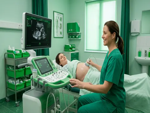
Comprehensive Diagnostic Sonography for Abdominal, Pelvic, Obstetric & Small-Parts Imaging
The Ultrasound Service at AS Medical Complex provides non-invasive diagnostic imaging for a wide range of medical conditions involving the abdomen, urinary system, pelvis, pregnancy, superficial organs, soft tissues, and selected blood vessels.
Ultrasound – also known as sonography – uses high-frequency sound waves to create real-time images of structures inside the body.
Unlike X-Ray and CT Scan, diagnostic ultrasound does not use ionizing radiation.
Ultrasound is commonly used because it can provide useful information about:
-
Internal organs
-
Fluid collections
-
Gallbladder
-
Liver
-
Kidneys
-
Urinary bladder
-
Pelvic organs
-
Pregnancy
-
Thyroid
-
Selected superficial structures
-
Selected blood vessels
-
Other clinically appropriate conditions
The Ali Medical reference Ultrasound service similarly identifies abdominal, pelvic, obstetric and small-parts imaging as core ultrasound applications.
At AS Medical Complex, the exact ultrasound examination should be selected according to the patient’s:
-
Symptoms
-
Medical history
-
Physical examination
-
Previous laboratory results
-
Previous imaging
-
Pregnancy status
-
Specialist recommendation
-
Clinical question
Ultrasound should not be performed simply because it is available. The examination should ideally answer a meaningful diagnostic question.
Our Ultrasound Services
Depending on clinical indication, available equipment, and appropriately trained personnel, Ultrasound services at AS Medical Complex may include:
-
Abdominal Ultrasound
-
Upper Abdominal Ultrasound
-
Whole Abdomen Ultrasound where appropriate
-
Liver Ultrasound
-
Gallbladder Ultrasound
-
Biliary-system assessment
-
Pancreatic assessment where adequately visualized
-
Spleen assessment
-
Kidney Ultrasound
-
Renal Ultrasound
-
Urinary Bladder Ultrasound
-
KUB-related ultrasound assessment
-
Assessment for hydronephrosis
-
Assessment for selected kidney stones
-
Pelvic Ultrasound
-
Female Pelvic Ultrasound
-
Transabdominal Pelvic Ultrasound
-
Transvaginal Ultrasound where clinically appropriate and available
-
Uterus assessment
-
Ovarian assessment
-
Fibroid assessment
-
Ovarian-cyst assessment
-
Obstetric Ultrasound
-
Early-pregnancy Ultrasound
-
Pregnancy viability assessment where clinically indicated
-
Pregnancy dating assessment
-
Fetal growth assessment where appropriate
-
Selected fetal-anatomy assessment where appropriate and available
-
Placental assessment where appropriate
-
Amniotic-fluid assessment where appropriate
-
Thyroid Ultrasound
-
Selected neck Ultrasound
-
Small-parts Ultrasound
-
Selected superficial soft-tissue Ultrasound
-
Scrotal/Testicular Ultrasound where available
-
Selected Doppler Ultrasound
-
Renal Doppler where clinically appropriate and available
-
Selected abdominal vascular Doppler
-
Selected obstetric Doppler
-
Selected superficial vascular assessment
-
Pediatric Ultrasound
-
Follow-up Ultrasound
-
Inpatient Ultrasound
-
Emergency Ultrasound where appropriate
-
Referral for specialized ultrasound where required
Specialized examinations should only be advertised as routine onsite services after confirming that the relevant equipment, transducers, reporting expertise, and trained personnel are available at AS Medical Complex.
What Is Ultrasound?
Ultrasound is a diagnostic imaging technique that uses sound waves to produce pictures of structures within the body.
During the examination, a device called a transducer sends high-frequency sound waves into the body.
These sound waves travel through tissues and reflect back toward the transducer.
A computer processes the returning echoes and creates images on a monitor.
Because the images are generated continuously, ultrasound can provide real-time imaging of many structures.
Ultrasound Does Not Use X-Ray Radiation
One of the major differences between ultrasound and imaging such as CT or X-Ray is that ultrasound does not use ionizing radiation.
Instead, it uses acoustic energy.
This makes ultrasound particularly useful in many situations where repeat imaging may be required and in pregnancy when ultrasound is medically indicated.
However, ultrasound should still be used responsibly.
FDA recommends applying the ALARA – As Low As Reasonably Achievable – principle, meaning exposure should be kept as low as reasonably possible while obtaining adequate diagnostic information.
Why Is Ultrasound Performed?
Ultrasound may be requested to investigate symptoms such as:
-
Abdominal pain
-
Pelvic pain
-
Abnormal vaginal bleeding
-
Urinary symptoms
-
Flank pain
-
Abnormal liver tests
-
Suspected gallstones
-
Abdominal swelling
-
Pregnancy-related concerns
-
Thyroid swelling
-
Scrotal pain
-
Superficial lumps
-
Other clinically appropriate symptoms
It may also be used to monitor known conditions or follow up abnormalities seen on another investigation.
Abdominal Ultrasound
An abdominal ultrasound produces images of structures within the abdomen.
It may examine organs such as:
-
Liver
-
Gallbladder
-
Bile ducts
-
Kidneys
-
Spleen
-
Pancreas where adequately visualized
-
Selected major blood vessels
-
Other accessible abdominal structures
RadiologyInfo describes abdominal ultrasound as an examination used to visualize organs and other structures within the upper abdomen.
Why Is Abdominal Ultrasound Performed?
Abdominal ultrasound may be considered in selected patients with:
-
Right upper abdominal pain
-
General abdominal pain
-
Abnormal liver-function tests
-
Suspected gallstones
-
Suspected gallbladder inflammation
-
Jaundice
-
Abdominal swelling
-
Suspected kidney abnormalities
-
Selected abdominal masses
-
Other abdominal conditions
The appropriate investigation may sometimes instead be:
-
CT Scan
-
MRI
-
MRCP
-
Endoscopy
-
Colonoscopy
-
Laboratory testing
depending on the suspected disease.
Liver Ultrasound
Ultrasound can provide information about the liver including:
-
Liver size
-
General appearance
-
Selected focal lesions
-
Fatty change
-
Biliary dilation
-
Other structural findings
However, ultrasound cannot always determine the exact cause or severity of liver disease.
Patients may require additional assessment such as:
-
Liver-function testing
-
Hepatitis testing
-
CT Scan
-
MRI
-
Gastroenterology consultation
-
Other investigations
Fatty Liver
Ultrasound may show features suggestive of fatty accumulation within the liver.
Fatty liver can be associated with factors such as:
-
Excess body weight
-
Diabetes
-
Metabolic disease
-
High triglycerides
-
Other medical conditions
An ultrasound finding alone does not determine the degree of liver inflammation or fibrosis.
Management may require coordination with:
-
Gastroenterology
-
General Medicine
-
Endocrinology
-
Dietitian Services
depending on the patient.
Liver Lesions
Ultrasound may identify selected liver abnormalities such as:
-
Cysts
-
Hemangioma-like lesions
-
Other focal abnormalities
Not every liver lesion represents cancer.
Depending on its appearance and the patient’s history, further evaluation may require:
-
Contrast-enhanced CT
-
MRI
-
Repeat ultrasound
-
Laboratory testing
-
Specialist assessment
Gallbladder Ultrasound
Ultrasound is commonly used to evaluate the gallbladder.
It may provide information about:
-
Gallstones
-
Gallbladder wall
-
Gallbladder distension
-
Fluid around the gallbladder
-
Biliary dilation
-
Other findings
Abdominal ultrasound is commonly used to evaluate gallstones and suspected gallbladder inflammation.
Gallstones
Gallstones are solid deposits that can form within the gallbladder.
Some patients have gallstones without any symptoms.
Others may develop symptoms such as:
-
Right upper abdominal pain
-
Pain after meals
-
Nausea
-
Vomiting
-
Pain radiating toward the back or shoulder
-
Other symptoms
Finding gallstones does not automatically mean surgery is required.
Treatment depends on:
-
Symptoms
-
Gallbladder inflammation
-
Bile-duct obstruction
-
Pancreatitis
-
Other complications
Acute Cholecystitis
Acute cholecystitis refers to inflammation of the gallbladder.
Possible symptoms include:
-
Persistent right upper abdominal pain
-
Fever
-
Nausea
-
Vomiting
-
Abdominal tenderness
Ultrasound can contribute important information, but diagnosis should be based on:
-
Symptoms
-
Examination
-
Laboratory tests
-
Imaging
Patients with significant symptoms may require General Surgery assessment.
Bile-Duct Abnormalities
Ultrasound may show dilation of the bile ducts.
Possible reasons include:
-
Bile-duct stones
-
Obstruction
-
Pancreatic or biliary disease
-
Other conditions
Ultrasound may not always identify the exact cause.
Further assessment may require:
-
MRCP
-
CT
-
Endoscopy
-
ERCP where clinically appropriate
-
Gastroenterology consultation
Pancreas Ultrasound
The pancreas may sometimes be visualized during abdominal ultrasound.
However, bowel gas and body anatomy can limit pancreatic imaging.
If the pancreas cannot be adequately assessed, the doctor may recommend:
-
CT Scan
-
MRI
-
MRCP
-
Other investigations
depending on the clinical question.
Spleen Ultrasound
Ultrasound may evaluate:
-
Spleen size
-
Selected structural abnormalities
-
Selected trauma-related findings
-
Other conditions
An enlarged spleen can occur in several medical conditions.
Further investigation depends on the suspected cause.
Abdominal Aortic Assessment
Ultrasound can be used to assess the abdominal aorta in selected patients, including evaluation for an abdominal aortic aneurysm.
RadiologyInfo includes abdominal aortic aneurysm among conditions that may be assessed with abdominal ultrasound.
A suspected ruptured aneurysm is an emergency and should not be managed through routine outpatient ultrasound.
Ascites
Ascites refers to abnormal fluid accumulation within the abdomen.
Ultrasound can help detect and localize abdominal fluid.
Possible causes include:
-
Liver disease
-
Heart disease
-
Cancer
-
Infection
-
Other medical conditions
The presence of fluid does not establish the cause.
Further medical evaluation is required.
Kidney Ultrasound
Renal ultrasound provides images of the kidneys and selected parts of the urinary tract.
It may be used to assess:
-
Kidney size
-
Kidney shape
-
Selected cysts or masses
-
Hydronephrosis
-
Urinary obstruction
-
Selected kidney stones
-
Other structural abnormalities
Renal Doppler may also provide information about renal blood flow in selected clinical situations.
Hydronephrosis
Hydronephrosis is dilation of the kidney’s urine-collecting system caused by impaired urine drainage.
Possible causes can include:
-
Kidney or ureteric stones
-
Enlarged prostate
-
Ureteric narrowing
-
Pelvic masses
-
Other urinary obstruction
Ultrasound may detect hydronephrosis, but another investigation may be needed to identify the exact cause.
Kidney Stones
Ultrasound can detect some kidney stones and signs of urinary obstruction.
However, it does not identify every stone equally well.
In selected adult patients with acute suspected renal colic, non-contrast CT may provide more detailed assessment.
Ultrasound can be particularly valuable when avoiding ionizing radiation is important, such as in selected:
-
Pregnant patients
-
Younger patients
-
Children
The correct imaging method depends on the clinical context.
Kidney Cysts
Simple kidney cysts are relatively common.
Ultrasound may help characterize whether a cyst appears simple or whether further imaging is needed.
A complex or uncertain renal lesion may require:
-
CT
-
MRI
-
Urology assessment
-
Nephrology assessment
depending on the findings.
Urinary Bladder Ultrasound
Bladder ultrasound may provide information about:
-
Bladder filling
-
Bladder wall
-
Urinary retention
-
Selected masses
-
Residual urine after urination
-
Other urinary abnormalities
A full bladder may be required for selected examinations.
Urinary Retention
Urinary retention occurs when the bladder cannot empty properly.
Symptoms may include:
-
Difficulty passing urine
-
Weak urinary stream
-
Lower abdominal discomfort
-
Inability to urinate
-
Overflow leakage
Ultrasound can help assess bladder distension and, where appropriate, residual urine after voiding.
Acute severe urinary retention may require urgent catheterization and Urology assessment.
Pelvic Ultrasound
Pelvic ultrasound uses sound waves to evaluate organs within the lower abdomen and pelvis.
RadiologyInfo describes three main pelvic ultrasound approaches:
-
Transabdominal
-
Transvaginal in women
-
Transrectal in selected male examinations
depending on the clinical indication.
At AS Medical Complex, availability of individual specialized pelvic techniques should be confirmed before booking.
Female Pelvic Ultrasound
Female pelvic ultrasound may evaluate structures such as:
-
Uterus
-
Endometrium
-
Ovaries
-
Cervix
-
Urinary bladder
-
Other pelvic structures
It may be recommended for selected patients with:
-
Pelvic pain
-
Abnormal vaginal bleeding
-
Heavy periods
-
Irregular periods
-
Suspected fibroids
-
Ovarian cysts
-
Pelvic masses
-
Fertility-related evaluation
-
Other gynecological symptoms
Pelvic ultrasound is commonly used to evaluate the uterus, ovaries, cervix and urinary bladder.
Transabdominal Pelvic Ultrasound
During transabdominal pelvic ultrasound, the transducer is moved over the lower abdomen.
A full urinary bladder is often required because it can improve visualization of pelvic structures.
Patients may therefore be instructed to drink water before the examination and avoid urinating until the scan is completed.
Transvaginal Ultrasound
A transvaginal ultrasound uses a specially designed ultrasound transducer inserted into the vagina.
It can provide more detailed images of selected pelvic structures than a transabdominal study.
It may be used for selected evaluation of:
-
Uterus
-
Endometrium
-
Ovaries
-
Early pregnancy
-
Pelvic pain
-
Abnormal bleeding
-
Other gynecological conditions
Transvaginal ultrasound should only be performed when clinically appropriate, with informed patient cooperation, privacy, appropriate infection-control procedures, and suitably trained staff.
Availability at AS Medical Complex should be confirmed before advertising it as a routine onsite service.
Fibroids
Uterine fibroids are non-cancerous growths arising from the muscular wall of the uterus.
Ultrasound can help assess:
-
Presence
-
Number
-
Size
-
Location
of fibroids.
Symptoms may include:
-
Heavy menstrual bleeding
-
Pelvic pressure
-
Pelvic pain
-
Urinary symptoms
-
Fertility-related issues in selected patients
Some fibroids cause no symptoms.
Treatment depends on symptoms rather than imaging alone.
Adenomyosis
Adenomyosis involves endometrial-type tissue within the muscular wall of the uterus.
Ultrasound may show findings suggestive of adenomyosis.
MRI may sometimes be recommended when the diagnosis remains uncertain or more detailed assessment is required.
Ovarian Cysts
Ultrasound can help assess ovarian cysts.
The scan may provide information about:
-
Size
-
Shape
-
Internal appearance
-
Simple or complex characteristics
-
Blood flow where Doppler is used
Many ovarian cysts are benign.
Management depends on:
-
Age
-
Menstrual status
-
Symptoms
-
Size
-
Ultrasound appearance
-
Persistence over time
Ovarian Masses
A complex ovarian mass may require further evaluation.
This can involve:
-
Repeat ultrasound
-
Doppler
-
Laboratory tests
-
MRI
-
CT
-
Gynecology consultation
-
Surgery in selected cases
An abnormal ovarian ultrasound does not automatically mean cancer.
Pelvic Pain
Pelvic pain can be caused by disorders involving:
-
Uterus
-
Ovaries
-
Urinary system
-
Gastrointestinal tract
-
Musculoskeletal system
Ultrasound may contribute to diagnosis, but not every cause of pelvic pain can be identified using ultrasound alone.
Abnormal Vaginal Bleeding
Pelvic ultrasound may be used as part of the evaluation of abnormal vaginal bleeding.
It can help assess:
-
Endometrial thickness
-
Fibroids
-
Polyps in selected situations
-
Ovarian abnormalities
-
Other uterine or pelvic findings
RadiologyInfo includes pelvic ultrasound among the imaging methods used in evaluation of abnormal vaginal bleeding.
Further assessment may sometimes require:
-
Gynecology examination
-
Laboratory testing
-
Endometrial sampling
-
Hysteroscopy
-
Other investigations
Obstetric Ultrasound
Obstetric ultrasound produces images of the developing embryo or fetus as well as maternal structures such as the uterus and ovaries.
It is an important imaging method in pregnancy because it does not use ionizing radiation.
Depending on gestational age and clinical indication, obstetric ultrasound may help assess:
-
Pregnancy location
-
Pregnancy dating
-
Fetal cardiac activity
-
Number of fetuses
-
Fetal growth
-
Placenta
-
Amniotic fluid
-
Selected fetal anatomy
-
Fetal position
-
Other pregnancy-related findings
Not every obstetric examination has the same purpose.
Early Pregnancy Ultrasound
An early-pregnancy ultrasound may be requested to assess:
-
Location of pregnancy
-
Gestational sac
-
Embryo where visible
-
Fetal cardiac activity where appropriate
-
Pregnancy dating
-
Vaginal bleeding
-
Pelvic pain
-
Other early-pregnancy concerns
Depending on gestational age, a transvaginal ultrasound may provide more information than a transabdominal examination.
Pregnancy Dating
Ultrasound can help estimate gestational age using appropriate fetal measurements.
Dating can be particularly useful when:
-
Menstrual dates are uncertain
-
Menstrual cycles are irregular
-
Pregnancy dating needs confirmation
-
There is a discrepancy between clinical dates and fetal size
The Obstetrics/Gynecology team should interpret dating together with the clinical history.
Pregnancy Viability
Ultrasound may help assess whether expected pregnancy structures and fetal cardiac activity are present according to gestational stage.
Very early pregnancy can sometimes be too early for a definitive conclusion.
In uncertain cases, the doctor may recommend:
-
Repeat ultrasound
-
Serial hCG testing
-
Clinical follow-up
A single early scan should not be interpreted without considering gestational timing.
Ectopic Pregnancy
An ectopic pregnancy occurs when a pregnancy develops outside the normal uterine cavity.
It can become life-threatening if rupture causes internal bleeding.
Warning symptoms may include:
-
Significant one-sided pelvic pain
-
Vaginal bleeding
-
Shoulder-tip pain
-
Dizziness
-
Fainting
-
Collapse
A patient with suspected ectopic pregnancy requires urgent medical and Gynecology assessment.
Ultrasound may be an important part of the evaluation, but it should not delay emergency treatment in an unstable patient.
Fetal Anatomy Ultrasound
At an appropriate stage of pregnancy, a detailed ultrasound may be used to assess aspects of fetal anatomy.
Such examinations can review structures including:
-
Brain
-
Spine
-
Heart
-
Abdomen
-
Limbs
-
Other structures
depending on the protocol and available expertise.
A normal ultrasound cannot guarantee that every fetal abnormality is absent.
Some abnormalities may:
-
Be too small to identify
-
Develop later
-
Be difficult to visualize
-
Require specialist fetal-medicine assessment
Specialized anomaly scanning should only be advertised if the relevant trained staff and protocols are available.
Fetal Growth Ultrasound
Ultrasound may be used to monitor fetal growth in selected pregnancies.
Measurements can help assess whether fetal size appears appropriate for gestational age.
Growth assessment may be particularly relevant in selected pregnancies involving:
-
Maternal hypertension
-
Diabetes
-
Previous fetal-growth problems
-
Other high-risk conditions
The Obstetrics team determines whether repeat growth scans are necessary.
Placenta
Obstetric ultrasound can help assess:
-
Placental location
-
Relationship to the cervix
-
Selected placental abnormalities
-
Other pregnancy-related concerns
Some placental conditions require repeat or specialist imaging later in pregnancy.
Amniotic Fluid
Ultrasound may also help estimate amniotic-fluid volume.
Too much or too little fluid can be associated with different pregnancy conditions and may require further Obstetric evaluation.
Multiple Pregnancy
Ultrasound is important in pregnancies involving more than one fetus.
It can help determine:
-
Number of fetuses
-
Pregnancy dating
-
Placental relationships
-
Growth
-
Other features
Multiple pregnancies may require closer specialist follow-up.
Doppler Ultrasound
Doppler ultrasound is a specialized ultrasound technique used to evaluate movement, particularly the movement of blood through blood vessels.
It can help doctors assess:
-
Direction of blood flow
-
Speed of blood flow
-
Narrowing of vessels
-
Blocked vessels
-
Selected organ blood supply
-
Other vascular conditions
RadiologyInfo describes Doppler as a technique used to evaluate blood flow through arteries and veins.
Types of Doppler
Depending on the equipment and examination, Doppler methods may include:
-
Color Doppler
-
Spectral Doppler
-
Power Doppler
These techniques provide different information about blood flow.
Availability depends on the ultrasound equipment and expertise available.
Renal Doppler
Selected patients may undergo renal Doppler to evaluate blood flow to the kidneys.
Possible indications may include assessment of:
-
Renal artery narrowing
-
Selected vascular abnormalities
-
Certain cases of difficult-to-control hypertension
-
Other renal vascular conditions
RadiologyInfo notes that renal Doppler can help assess renal blood flow and selected narrowing or clotting within renal vessels.
Obstetric Doppler
Doppler may form part of selected obstetric ultrasound examinations to evaluate blood flow involving:
-
Umbilical cord
-
Placenta
-
Fetus
RadiologyInfo notes that Doppler can be incorporated into obstetric ultrasound when clinically appropriate.
Doppler should be used for a valid medical indication and with appropriate attention to ultrasound exposure.
Doppler Safety in Pregnancy
Ultrasound exposure should be kept as low as reasonably achievable.
This is especially important when Doppler techniques are used in pregnancy.
AIUM recommends prudent use of diagnostic ultrasound during pregnancy and minimizing exposure duration and output while obtaining the required medical information.
Thyroid Ultrasound
Thyroid ultrasound provides images of the thyroid gland and surrounding superficial structures.
It may be used for selected patients with:
-
Thyroid enlargement
-
Thyroid nodules
-
Neck swelling
-
Abnormal physical examination
-
Follow-up of known thyroid lesions
-
Other appropriate indications
Ultrasound may provide information about:
-
Nodule size
-
Shape
-
Composition
-
Other imaging features
A thyroid ultrasound cannot by itself determine whether every nodule is benign or malignant.
Thyroid Nodules
Thyroid nodules are common.
Many are benign.
Depending on ultrasound appearance and clinical factors, further evaluation may involve:
-
Thyroid-function tests
-
Follow-up ultrasound
-
Fine-needle aspiration
-
Endocrinology consultation
-
Surgery in selected patients
The management plan should consider both imaging and clinical risk.
Small-Parts Ultrasound
The Ali Medical source specifically identifies small-parts imaging as part of its Ultrasound service.
Small-parts ultrasound may include selected assessment of superficial structures such as:
-
Thyroid
-
Scrotum
-
Selected soft tissues
-
Superficial lumps
-
Other accessible structures
depending on the equipment and expertise available at AS Medical Complex.
Scrotal & Testicular Ultrasound
Scrotal ultrasound can assess structures such as:
-
Testicles
-
Epididymis
-
Scrotal tissues
-
Selected blood flow with Doppler
Possible reasons include:
-
Testicular pain
-
Swelling
-
Lump
-
Trauma
-
Suspected infection
-
Other abnormalities
RadiologyInfo notes that ultrasound is useful in evaluating scrotal pain, including in pediatric patients.
Sudden Testicular Pain
Sudden severe testicular pain can represent testicular torsion, where blood flow to the testicle becomes compromised.
This is an emergency.
A patient with sudden severe testicular pain should seek urgent medical assessment rather than waiting for a routine ultrasound appointment.
Doppler ultrasound may assist diagnosis, but emergency Urology or surgical assessment should not be unnecessarily delayed.
Soft-Tissue Ultrasound
Ultrasound may help assess selected superficial abnormalities such as:
-
Cysts
-
Lipoma-like masses
-
Abscesses
-
Fluid collections
-
Other superficial lesions
Ultrasound can help determine whether an abnormality appears:
-
Solid
-
Fluid-filled
-
Mixed
Further assessment may sometimes require:
-
MRI
-
CT
-
Biopsy
-
Surgery
-
Specialist consultation
Abscess
An abscess is a collection of infected fluid.
Possible symptoms include:
-
Pain
-
Swelling
-
Redness
-
Warmth
-
Fever
Ultrasound can sometimes help confirm whether a superficial swelling contains fluid.
An abscess may require:
-
Drainage
-
Antibiotics
-
Surgical assessment
depending on the clinical situation.
Pediatric Ultrasound
Ultrasound can be particularly useful in children because it does not expose them to ionizing radiation.
Pediatric ultrasound may help evaluate selected:
-
Abdominal pain
-
Pelvic pain
-
Urinary conditions
-
Scrotal pain
-
Other appropriate problems
RadiologyInfo notes that pediatric ultrasound is particularly useful for abdominal, pelvic and scrotal assessment.
The appropriate examination should be selected according to the child’s age, symptoms and clinical condition.
Ultrasound-Guided Procedures
Ultrasound can provide real-time guidance during selected procedures.
Possible applications can include:
-
Needle placement
-
Fluid aspiration
-
Biopsy
-
Drainage
-
Selected vascular-access procedures
RadiologyInfo notes that ultrasound may be used to guide procedures such as needle biopsies.
Such procedures require:
-
Trained clinicians
-
Appropriate consent
-
Sterile technique
-
Suitable equipment
-
Monitoring where required
They should only be advertised as AS Medical Complex services after onsite capability is confirmed.
Ultrasound Preparation
Preparation depends on the body area being examined.
Some ultrasound examinations require:
-
Fasting
-
A full urinary bladder
-
No special preparation
-
Other specific instructions
Patients should follow the instructions provided when the examination is booked.
Fasting for Abdominal Ultrasound
Patients undergoing selected abdominal ultrasound examinations may be asked to avoid food for a period before the scan.
Fasting can help improve visualization of structures such as:
-
Gallbladder
-
Upper abdominal organs
and may reduce some limitations caused by bowel contents.
Preparation should follow the instructions given for the specific examination.
Full Bladder for Pelvic Ultrasound
A full urinary bladder can improve transabdominal visualization of the pelvis.
Patients may therefore be asked to:
-
Drink water before the examination
-
Avoid urinating until the scan is complete
RadiologyInfo specifically notes that preparation for pelvic ultrasound may involve drinking water to fill the bladder.
Medicines Before Ultrasound
Most routine ultrasound examinations do not require patients to stop their regular medications.
However, patients should follow any specific preparation instructions provided by:
-
Radiology
-
Treating doctor
-
Obstetrics/Gynecology team
Patients should not independently stop prescribed medicines solely because an ultrasound has been scheduled.
What Should You Wear?
Patients should wear comfortable clothing that allows the body area being examined to be accessed.
Depending on the examination, the patient may be asked to:
-
Lift or adjust clothing
-
Remove selected clothing
-
Change into a hospital gown
Privacy should be maintained throughout the examination.
What Happens During an Ultrasound?
During a typical external ultrasound:
-
Patient identity is confirmed.
-
The clinical indication is reviewed.
-
The patient lies on an examination table.
-
The relevant body area is exposed.
-
Ultrasound gel is placed on the skin.
-
The transducer is moved over the area.
-
Real-time images are obtained.
-
Measurements may be taken.
-
Doppler may be used if required.
The examination is generally non-invasive and well tolerated.
Ultrasound Gel
A water-based gel is applied between the skin and transducer.
The gel removes air between the probe and skin, helping sound waves travel efficiently into the body.
The gel is wiped away after the examination.
Is Ultrasound Painful?
Most routine ultrasound examinations are painless.
Some patients may feel mild discomfort when the transducer presses over an area that is already:
-
Tender
-
Inflamed
-
Injured
Internal pelvic ultrasound may also cause temporary discomfort in selected patients.
Patients should tell the person performing the examination if they experience significant pain.
How Long Does an Ultrasound Take?
The duration depends on:
-
Body area
-
Complexity
-
Number of organs being examined
-
Need for Doppler
-
Patient cooperation
-
Image quality
-
Specialized measurements
A focused study may be relatively short, while a comprehensive examination may take longer.
AS Medical Complex should therefore avoid promising one fixed time for every ultrasound.
Ultrasound Results
Ultrasound images are reviewed by an appropriately qualified radiologist or imaging professional according to the hospital’s reporting pathway.
The report may describe:
-
Normal findings
-
Abnormal findings
-
Organ size
-
Masses
-
Cysts
-
Stones
-
Fluid
-
Blood-flow findings
-
Other observations
The report should be interpreted together with:
-
Symptoms
-
Examination
-
Laboratory results
-
Previous imaging
Ultrasound Image vs Ultrasound Report
The images show the anatomy captured during the examination.
The report provides the professional interpretation.
Patients should avoid trying to diagnose themselves solely by looking at individual ultrasound images.
A finding may require clinical context before its significance can be understood.
Incidental Findings
Ultrasound occasionally identifies an unexpected abnormality unrelated to the original reason for testing.
Possible next steps may include:
-
No further action
-
Follow-up ultrasound
-
CT
-
MRI
-
Laboratory testing
-
Specialist consultation
Not every incidental abnormality represents serious disease.
Ultrasound Limitations
Ultrasound is extremely useful but has limitations.
Image quality can be reduced by:
-
Bowel gas
-
Obesity
-
Deep location of an organ
-
Bone
-
Air-filled lung
-
Wounds
-
Dressings
-
Patient movement
-
Limited acoustic windows
Ultrasound does not pass effectively through bone and air, limiting evaluation of some structures.
Another imaging method may therefore be required.
A Normal Ultrasound Does Not Exclude Every Disease
A normal ultrasound does not guarantee that all disease is absent.
Some abnormalities:
-
Are too small
-
Are hidden by bowel gas
-
Require CT
-
Require MRI
-
Require endoscopy
-
Require laboratory testing
-
Require biopsy
The doctor should decide whether further investigation is needed if symptoms continue.
Ultrasound vs CT Scan
Ultrasound
Advantages can include:
-
No ionizing radiation
-
Real-time imaging
-
Good evaluation of many abdominal and pelvic structures
-
Useful pregnancy imaging
-
Relatively quick assessment
-
Ability to assess blood flow with Doppler
CT Scan
CT may provide more detailed evaluation of:
-
Major trauma
-
Deep abdominal structures
-
Lungs
-
Complex bowel disease
-
Kidney stones
-
Internal bleeding
-
Many emergencies
CT uses ionizing radiation.
The appropriate examination depends on the clinical question.
Ultrasound vs MRI
MRI may provide more detailed evaluation of selected:
-
Brain
-
Spinal cord
-
Nerves
-
Deep soft tissues
-
Complex pelvic conditions
-
Musculoskeletal structures
-
Certain liver or pancreatic abnormalities
MRI does not use ionizing radiation but generally takes longer and has different safety considerations.
Ultrasound and MRI therefore complement rather than replace one another.
Ultrasound vs X-Ray
X-Ray is particularly useful for:
-
Bone fractures
-
Joint alignment
-
Chest conditions
-
Other radiographic indications
Ultrasound is more useful for many:
-
Soft tissues
-
Abdominal organs
-
Pelvic organs
-
Pregnancy-related assessments
-
Fluid collections
The doctor should select the appropriate method.
Ultrasound vs Portable Ultrasound
AS Medical Complex may maintain separate pathways for:
Departmental Ultrasound
Designed for more comprehensive standardized imaging in the Radiology Department.
Portable Ultrasound
Designed for selected bedside examinations when moving the patient is difficult or when focused imaging is clinically useful.
A portable focused examination may not always replace a comprehensive departmental ultrasound.
Ultrasound Safety
Diagnostic ultrasound has been used clinically for decades.
AIUM states that no independently confirmed harmful effects have been demonstrated in human patients from appropriately used present-day diagnostic ultrasound equipment in the absence of contrast agents.
However, ultrasound does deposit acoustic energy within tissue.
For this reason, FDA recommends:
-
Appropriate clinical use
-
Limiting exposure duration
-
Using only the output needed for diagnostic-quality imaging
-
Following ALARA principles
Ultrasound During Pregnancy
Ultrasound is the preferred imaging method for many pregnancy-related assessments because it does not use ionizing radiation.
However, pregnancy scanning should be performed for a medical purpose and by appropriately trained healthcare professionals.
AIUM discourages unnecessary nonmedical fetal ultrasound examinations and recommends prudent use.
Keepsake Fetal Ultrasound
Ultrasound should not be prolonged or performed solely for entertainment or nonmedical fetal imaging.
The purpose of hospital ultrasound should remain:
-
Diagnosis
-
Pregnancy assessment
-
Monitoring
-
Clinical decision-making
rather than obtaining unnecessary souvenir images.
Infection Prevention
Ultrasound equipment and transducers come into direct contact with patients.
Appropriate infection-prevention procedures should therefore be followed.
This can include:
-
Cleaning
-
Disinfection
-
Appropriate probe covers where required
-
Hand hygiene
-
Safe gel practices
Internal ultrasound probes require higher-level infection-control precautions than routine external probes.
When Should You Consider an Ultrasound?
You may require Ultrasound assessment if your doctor is investigating:
-
Abdominal pain
-
Gallstones
-
Gallbladder inflammation
-
Abnormal liver tests
-
Liver abnormalities
-
Kidney abnormalities
-
Hydronephrosis
-
Urinary retention
-
Pelvic pain
-
Abnormal vaginal bleeding
-
Fibroids
-
Ovarian cysts
-
Pregnancy-related concerns
-
Thyroid nodules
-
Superficial lumps
-
Scrotal symptoms
-
Other clinically appropriate conditions
The examination should be targeted to the medical question.
When Should You See a Doctor Before Booking Ultrasound?
Medical assessment may be appropriate first when symptoms are unclear.
For example:
-
General abdominal discomfort
-
Unexplained weight loss
-
Persistent fever
-
Severe fatigue
-
Recurrent vomiting
-
Persistent urinary symptoms
-
Abnormal bleeding
A doctor can determine whether the best investigation is:
-
Ultrasound
-
CT
-
MRI
-
X-Ray
-
Laboratory testing
-
Endoscopy
-
Another examination
When Is Emergency Care Required?
Do not wait for a routine ultrasound appointment if you develop:
-
Severe abdominal pain with collapse
-
Suspected ectopic pregnancy with severe pain or bleeding
-
Sudden severe testicular pain
-
Major trauma
-
Severe internal or external bleeding
-
Loss of consciousness
-
Severe breathing difficulty
-
Shock
-
Rapidly worsening infection
-
Other potentially life-threatening symptoms
These situations require urgent clinical assessment.
Ultrasound may form part of Emergency care, but the patient may also require:
-
CT Scan
-
X-Ray
-
Laboratory testing
-
Surgery
-
Other urgent treatment
Multidisciplinary Ultrasound Care
Depending on the ultrasound findings, care may be coordinated with:
-
Radiology
-
General Medicine
-
Gastroenterology
-
General & Laparoscopic Surgery
-
Gynecology
-
Urology
-
Nephrology
-
Endocrinology
-
Pediatrics
-
Emergency Services
-
Vascular Surgery
-
Pulmonology
-
Other relevant specialties
Ultrasound is therefore frequently one part of a larger diagnostic and treatment pathway.
Follow-Up After Ultrasound
The next step depends on:
-
Ultrasound findings
-
Patient symptoms
-
Laboratory results
-
Medical history
-
Specialist assessment
Possible next steps may include:
-
No further investigation
-
Medication
-
Follow-up ultrasound
-
CT Scan
-
MRI
-
MRCP
-
Laboratory testing
-
Endoscopy
-
Colonoscopy
-
Biopsy
-
Surgery
-
Specialist consultation
Significant findings should be reviewed with the healthcare professional responsible for the patient’s care.
Repeat Ultrasound
Repeat ultrasound may be appropriate for selected conditions such as:
-
Pregnancy monitoring
-
Ovarian cyst follow-up
-
Liver-lesion surveillance
-
Kidney abnormalities
-
Thyroid nodules
-
Other clinically appropriate conditions
Because ultrasound does not use ionizing radiation, repeat examinations can be performed when medically indicated.
However, repeated imaging should still have a clear clinical purpose and follow prudent ultrasound-use principles.
Ultrasound FAQs
What is an Ultrasound?
Ultrasound, or sonography, is a diagnostic imaging technique that uses high-frequency sound waves to create real-time pictures of structures inside the body. It can examine organs, soft tissues, pregnancy-related structures and selected blood vessels without using ionizing radiation.
Is Ultrasound safe?
Diagnostic ultrasound is generally considered safe when used appropriately. It does not use ionizing radiation. However, ultrasound introduces acoustic energy into tissue, so FDA recommends using the lowest output and shortest exposure needed to obtain useful diagnostic information according to the ALARA principle.
Do I need to fast before an Ultrasound?
It depends on the examination. Selected abdominal ultrasound studies may require fasting, while transabdominal pelvic ultrasound may require a full urinary bladder. Many other ultrasound examinations require little or no preparation. Follow the instructions provided for your specific scan.
Can Ultrasound be performed during pregnancy?
Yes. Ultrasound is widely used for medically indicated pregnancy assessment because it does not use ionizing radiation. It can provide information about the embryo or fetus, uterus, ovaries, placenta and other pregnancy-related structures. Pregnancy ultrasound should nevertheless be used prudently for medical purposes rather than unnecessary nonmedical scanning.
Can Ultrasound detect kidney stones and gallstones?
Ultrasound is commonly used to detect gallstones and may detect selected kidney stones or signs of urinary obstruction. However, it does not identify every kidney stone, and CT may be more appropriate in some patients. The correct examination depends on symptoms and clinical circumstances.
What is the difference between Ultrasound and Doppler Ultrasound?
Routine ultrasound mainly creates images of body structures. Doppler ultrasound is a specialized technique that evaluates movement, particularly blood flow through arteries and veins. Doppler may be added when doctors need information about blood flow to an organ, fetus, or blood vessel.
What happens if my Ultrasound is normal but I still have symptoms?
A normal ultrasound does not exclude every medical condition. Bowel gas, body anatomy, depth, bone and air can limit ultrasound visualization, and some diseases are better assessed using CT, MRI, endoscopy, laboratory testing or another investigation. Persistent or worsening symptoms should therefore be reviewed by the treating doctor.
Ultrasound Services at AS Medical Complex
The Ultrasound Service at AS Medical Complex is designed to provide safe, non-invasive and clinically appropriate diagnostic imaging for patients requiring assessment of the abdomen, kidneys, urinary tract, pelvis, pregnancy, thyroid, superficial structures, and other medically indicated areas.
From abdominal, liver, gallbladder, kidney, urinary-bladder and pelvic ultrasound to obstetric imaging, selected small-parts examinations, thyroid assessment and Doppler studies where appropriate and available, our goal is to provide diagnostic information that supports effective medical decision-making.
Ultrasound has the important advantage of providing real-time imaging without ionizing radiation, but it also has limitations. Some medical conditions require CT Scan, MRI, X-Ray, laboratory testing, endoscopy, biopsy, or specialist assessment for complete evaluation.
Patient safety and diagnostic quality remain central to every examination. Ultrasound should be performed for an appropriate medical indication, with acoustic exposure kept as low as reasonably achievable while obtaining the information necessary for diagnosis.
Ultrasound findings can be coordinated with the relevant departments at AS Medical Complex, including Radiology, Gastroenterology, General & Laparoscopic Surgery, Gynecology, Urology, Nephrology, Endocrinology, Pediatrics, Emergency Services, and other specialties according to clinical need.
Book an Ultrasound at AS Medical Complex for physician-requested abdominal, kidney, urinary, pelvic, obstetric, thyroid, small-parts, Doppler, and other clinically indicated ultrasound examinations.