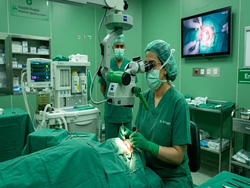
Ophthalmology Surgery at AS Medical Complex
Contact hospital
- Availability
Contact hospital
- Booking
WhatsApp / call
- Location
Islamabad
Comprehensive Eye, Vision & Ophthalmic Care
The Ophthalmology Department at AS Medical Complex provides comprehensive assessment, diagnosis, medical treatment, surgical consultation, and ongoing care for conditions affecting the eyes and vision.
Eye conditions can involve the eyelids, cornea, lens, retina, optic nerve, eye pressure, tear system, eye muscles, or other structures responsible for vision.
Some eye disorders produce noticeable symptoms such as blurred vision, pain, redness, or flashes and floaters, while others—particularly conditions such as glaucoma and early diabetic retinopathy—may develop without obvious symptoms.
The Ali Medical reference Ophthalmology department similarly provides routine eye examinations, vision screening, and treatment for conditions including cataracts, glaucoma, macular degeneration, and diabetic eye disease.
At AS Medical Complex, our aim is to support early diagnosis, preservation of vision, appropriate medical or surgical treatment, and long-term eye health.
Our Ophthalmology Services
The Ophthalmology Department at AS Medical Complex may provide assessment and management for:
-
Comprehensive eye examinations
-
Vision testing
-
Blurred or reduced vision
-
Refractive errors
-
Myopia – nearsightedness
-
Hyperopia – farsightedness
-
Astigmatism
-
Presbyopia
-
Cataracts
-
Cataract surgical assessment
-
Glaucoma
-
Raised eye pressure
-
Diabetic eye disease
-
Diabetic retinopathy
-
Macular edema
-
Age-related macular degeneration
-
Retinal disorders
-
Retinal tears and retinal detachment
-
Flashes and floaters
-
Dry eye disease
-
Corneal diseases
-
Keratitis
-
Eye allergies
-
Conjunctivitis
-
Red eye
-
Blepharitis
-
Eyelid disorders
-
Styes and chalazia
-
Uveitis
-
Eye infections
-
Eye trauma
-
Foreign body in the eye
-
Pediatric eye conditions
-
Amblyopia – lazy eye
-
Squint / strabismus assessment
-
Vision problems in children
-
Ophthalmic complications of systemic disease
-
Eye examination for patients with diabetes
-
Ophthalmic diagnostic testing
-
Laser or injection-based retinal treatment where clinically indicated and available
-
Ophthalmic surgical consultation
-
Postoperative eye care
-
Emergency ophthalmic assessment
The exact investigations or treatment required depend on the patient's symptoms, eye examination, age, medical history, and diagnosis.
Comprehensive Eye Examination
Routine Eye & Vision Assessment
A comprehensive eye examination can help assess both how clearly a patient sees and whether important structures inside the eye are healthy.
Depending on the patient's age and symptoms, an examination may involve:
-
Visual-acuity testing
-
Refraction
-
Eye-pressure measurement
-
Slit-lamp examination
-
Examination of the cornea
-
Examination of the lens
-
Pupil assessment
-
Eye-movement assessment
-
Dilated retinal examination
-
Optic-nerve assessment
-
Additional diagnostic tests where required
Regular examinations can be particularly important for patients with:
-
Diabetes
-
Glaucoma risk
-
Family history of eye disease
-
Previous eye surgery
-
Significant refractive error
-
Increasing age
-
Chronic medical conditions affecting the eyes
The National Eye Institute emphasizes comprehensive dilated eye examinations for detecting several important eye diseases, some of which may have few or no early symptoms.
Refractive Errors & Blurred Vision
Refractive Errors
A refractive error occurs when the shape or focusing system of the eye does not focus light accurately on the retina.
The major types are:
-
Myopia
-
Hyperopia
-
Astigmatism
-
Presbyopia
Blurred vision is the most common symptom. Patients may also experience headaches, squinting, eye strain, glare, or difficulty focusing while reading or using screens.
Treatment may involve:
-
Eyeglasses
-
Contact lenses
-
Other vision-correction options where appropriate
Myopia – Nearsightedness
Myopia causes distant objects to appear blurred while nearer objects may remain clearer.
It commonly begins during childhood or adolescence.
Patients may:
-
Squint to see distant objects
-
Have difficulty seeing a classroom board
-
Move closer to televisions or screens
-
Experience eye strain
-
Require progressively updated glasses
High myopia can also increase the risk of certain retinal complications and therefore may require more detailed eye monitoring.
Hyperopia – Farsightedness
Hyperopia occurs when the eye focuses light behind rather than directly on the retina.
Depending on age and severity, patients may experience:
-
Difficulty with near work
-
Eye strain
-
Headache after reading
-
Blurred near vision
-
Sometimes blurred distance vision
An eye examination can determine whether corrective lenses are appropriate.
Astigmatism
Astigmatism results from differences in the curvature or shape of the cornea or lens.
Patients may experience:
-
Blurred vision
-
Distorted vision
-
Eye strain
-
Headaches
-
Difficulty seeing clearly at night
Astigmatism can occur together with myopia or hyperopia.
Presbyopia
Presbyopia is an age-related reduction in the eye's ability to focus clearly on nearby objects.
It commonly becomes noticeable in middle age.
Patients may:
-
Hold reading material farther away
-
Need more light to read
-
Experience eye strain during close work
-
Have difficulty reading small print
NEI notes that presbyopia occurs as the natural lens becomes less flexible with age.
Cataract
What Is a Cataract?
A cataract is clouding of the eye's natural lens.
As the lens becomes cloudy, light cannot pass through and focus as clearly as before.
Patients may notice:
-
Blurred vision
-
Hazy vision
-
Reduced contrast
-
Glare
-
Halos around lights
-
Difficulty driving at night
-
Colors appearing less vivid
-
Frequent changes in glasses prescription
The National Eye Institute defines a cataract as a cloudy area in the eye's lens and notes that cataracts become increasingly common with age.
Causes & Risk Factors for Cataract
Age-related change is one of the most common causes.
Other factors can include:
-
Diabetes
-
Eye injury
-
Previous eye surgery
-
Long-term use of certain medications
-
Some congenital conditions
-
Other eye diseases
A cataract may affect one or both eyes.
Cataract Assessment
Assessment may include:
-
Visual-acuity testing
-
Refraction
-
Slit-lamp examination
-
Dilated eye examination
-
Eye-pressure measurement
-
Retinal assessment
-
Additional measurements when surgery is planned
The ophthalmologist also considers whether another eye condition—such as glaucoma, retinal disease, or macular degeneration—is contributing to reduced vision.
Cataract Treatment
Early cataracts may sometimes be managed with:
-
Updated glasses
-
Improved lighting
-
Adjustments to daily activities
When cataract significantly interferes with everyday vision, cataract surgery may be considered.
Surgery is the definitive treatment used to remove a cataract.
Cataract Surgery
During cataract surgery, the cloudy natural lens is removed and usually replaced with an artificial intraocular lens (IOL).
Surgical planning typically considers:
-
Degree of cataract
-
Current vision
-
Eye measurements
-
Corneal health
-
Retina
-
Optic nerve
-
Existing eye diseases
-
Patient's visual needs
-
Overall medical condition
The exact surgical technique and lens choice should be discussed with the treating ophthalmologist.
After Cataract Surgery
Postoperative care may include:
-
Prescribed eye drops
-
Avoiding rubbing the eye
-
Protection from contamination or injury
-
Temporary activity restrictions
-
Follow-up examination
-
Monitoring eye pressure
-
Monitoring healing
Patients should seek prompt review if they develop significant pain, sudden loss of vision, increasing redness, flashes, or a sudden increase in floaters after surgery.
Glaucoma
What Is Glaucoma?
Glaucoma refers to a group of eye diseases that damage the optic nerve, which carries visual information from the eye to the brain.
Glaucoma can gradually cause permanent loss of vision.
Importantly, many forms of glaucoma develop without obvious early symptoms. NEI emphasizes that comprehensive dilated eye examination is important for detecting the disease.
Eye Pressure & Glaucoma
Raised pressure inside the eye is an important glaucoma risk factor, although glaucoma can sometimes develop even when measured eye pressure is within a typical range.
NEI notes that higher intraocular pressure increases glaucoma risk and that lowering eye pressure can help prevent further vision loss in affected patients.
Glaucoma Assessment
Evaluation may include:
-
Eye-pressure measurement
-
Optic-nerve examination
-
Dilated eye examination
-
Visual-field testing
-
Optical Coherence Tomography – OCT
-
Corneal-thickness measurement where appropriate
-
Gonioscopy
-
Fundus photography
-
Other investigations
Not every patient requires all tests during every visit.
Who Should Be Particularly Alert for Glaucoma?
Risk may be higher in people with:
-
Increasing age
-
Family history of glaucoma
-
Raised intraocular pressure
-
Certain eye conditions
-
Previous eye injury
-
Long-term use of some medications
-
Other individual risk factors
Because early glaucoma may be silent, people at higher risk should discuss appropriate examination intervals with an eye specialist.
Glaucoma Treatment
Treatment aims to preserve remaining vision by preventing or slowing further optic-nerve damage.
Depending on the condition, treatment may involve:
-
Prescription eye drops
-
Laser treatment
-
Surgery
-
Combination treatment
Treatment cannot normally restore vision already permanently lost from glaucoma, making early diagnosis particularly important.
Acute Angle-Closure Glaucoma
Some forms of glaucoma can cause a sudden dangerous rise in eye pressure.
Possible symptoms include:
-
Severe eye pain
-
Red eye
-
Sudden blurred vision
-
Halos around lights
-
Headache
-
Nausea
-
Vomiting
This requires urgent ophthalmic assessment and should not wait for a routine appointment.
Diabetic Eye Disease
Diabetes & Eye Health
Diabetes can affect several structures of the eye and increase the risk of serious vision problems.
Diabetic eye disease includes conditions such as:
-
Diabetic retinopathy
-
Diabetic macular edema
-
Cataract
-
Glaucoma
NEI recommends regular comprehensive dilated eye examinations for people with diabetes because diabetic retinopathy may initially cause no symptoms.
Diabetic Retinopathy
Diabetic retinopathy occurs when diabetes damages blood vessels in the retina.
The retina is the light-sensitive tissue at the back of the eye.
Early disease may produce no noticeable symptoms.
As the condition progresses, patients may develop:
-
Blurred vision
-
Floaters
-
Dark areas in vision
-
Difficulty seeing clearly
-
Progressive vision loss
Diabetic retinopathy can cause severe visual impairment if advanced disease is not appropriately managed.
Diabetic Eye Examination
A diabetic eye assessment may include:
-
Visual-acuity testing
-
Dilated retinal examination
-
Eye-pressure measurement
-
Retinal photography
-
OCT
-
Additional retinal imaging or angiography when appropriate
Follow-up frequency depends on the retinal findings, pregnancy status where relevant, diabetes control, and other clinical factors.
Diabetic Macular Edema
Diabetic macular edema (DME) occurs when fluid accumulates within the macula because damaged retinal blood vessels leak.
The macula is responsible for detailed central vision.
Patients may experience:
-
Blurred central vision
-
Distorted vision
-
Difficulty reading
-
Reduced visual detail
Treatment may involve retinal injections or other specialist retinal treatment depending on the condition.
Intravitreal Injections
Certain retinal diseases can be treated using medicines injected into the eye.
Anti-VEGF injections may be used for selected conditions including:
-
Diabetic retinopathy
-
Diabetic macular edema
-
Macular edema caused by retinal vascular disease
-
Wet age-related macular degeneration
NEI describes anti-VEGF injections as an established treatment for several retinal conditions involving abnormal or leaking blood vessels.
Such treatment should only be provided after appropriate retinal assessment.
Retina
Retinal Disorders
The retina converts light entering the eye into signals that are transmitted toward the brain.
Retinal disorders can therefore significantly affect vision.
The Ophthalmology Department may assess patients with:
-
Diabetic retinopathy
-
Macular edema
-
Retinal tears
-
Retinal detachment
-
Retinal vascular disorders
-
Age-related macular degeneration
-
Macular problems
-
Vitreous disorders
-
Other retinal abnormalities
Flashes & Floaters
Floaters may appear as:
-
Small spots
-
Lines
-
Cobweb-like shapes
-
Moving shadows
Flashes may appear as brief flashes or streaks of light.
Many floaters are related to age-related changes within the vitreous gel, but sudden new symptoms may sometimes indicate a retinal tear.
Urgent eye assessment is particularly important when there is:
-
Sudden onset of many new floaters
-
Repeated flashes
-
A shadow in the vision
-
A curtain-like area of vision loss
-
Sudden reduction in vision
Retinal Tear
A retinal tear occurs when a break forms in the retina.
A tear may sometimes progress to retinal detachment.
The American Academy of Ophthalmology identifies a retinal tear as a serious condition requiring ophthalmic assessment.
Selected retinal tears can be treated with laser or other procedures to reduce the risk of progression.
Retinal Detachment
Retinal detachment occurs when the retina separates from its normal position at the back of the eye.
Warning symptoms can include:
-
Sudden increase in floaters
-
Flashing lights
-
Shadow in peripheral vision
-
Curtain-like loss of vision
-
Sudden visual deterioration
Retinal detachment is an ophthalmic emergency.
Prompt diagnosis and treatment are important to preserve as much vision as possible.
Vitrectomy
Vitrectomy is an eye operation used for selected problems involving the retina and vitreous gel.
Depending on the diagnosis, it may be considered for conditions such as:
-
Certain retinal detachments
-
Vitreous hemorrhage
-
Selected diabetic retinal complications
-
Macular conditions
-
Other vitreoretinal disorders
AAO describes vitrectomy as surgery used for problems involving the retina and vitreous.
Availability of vitreoretinal surgery at AS Medical Complex should be determined according to specialist and hospital facilities.
Age-Related Macular Degeneration
AMD
Age-Related Macular Degeneration (AMD) affects the macula, the central part of the retina responsible for sharp detailed vision.
It may make activities such as:
-
Reading
-
Recognizing faces
-
Driving
-
Detailed close work
more difficult.
NEI describes AMD as a disease that blurs central vision and is an important cause of vision loss among older adults.
Dry & Wet AMD
AMD can occur in different forms and stages.
Dry AMD is generally associated with gradual degenerative changes.
Wet AMD involves abnormal blood vessels that can leak fluid or blood into the retina and may cause more rapid central visual loss.
Patients may notice:
-
Blurred central vision
-
Distortion
-
Straight lines appearing wavy
-
Dark or blank areas in central vision
Treatment of Wet AMD
Anti-VEGF injections are commonly used to treat wet AMD by reducing abnormal vascular leakage and growth.
NEI identifies anti-VEGF injection treatment as a major treatment for wet AMD.
Treatment frequency and monitoring depend on the individual eye and response to therapy.
Dry Eye Disease
Dry Eyes
Dry eye disease occurs when the eyes do not produce enough tears or when the tears do not adequately protect and lubricate the eye surface.
Symptoms can include:
-
Burning
-
Stinging
-
Gritty sensation
-
Feeling that something is in the eye
-
Redness
-
Watering
-
Light sensitivity
-
Intermittent blurred vision
-
Eye fatigue
Causes of Dry Eye
Possible contributing factors include:
-
Reduced tear production
-
Excessive tear evaporation
-
Eyelid-gland problems
-
Contact-lens use
-
Increasing age
-
Certain medications
-
Autoimmune disease
-
Long periods of screen use
-
Environmental factors
Treatment should be directed toward the underlying cause where possible.
Dry Eye Treatment
Depending on severity, management may include:
-
Artificial tears
-
Lubricating gels or ointments
-
Eyelid hygiene
-
Environmental modification
-
Prescription medicines in selected patients
-
Treatment of associated eyelid disease
-
Other specialist treatment
NEI notes that treatment varies according to the cause and severity of dry eye.
Cornea
Corneal Conditions
The cornea is the clear front surface of the eye.
Conditions affecting it may cause:
-
Pain
-
Redness
-
Light sensitivity
-
Watering
-
Blurred vision
-
Foreign-body sensation
Corneal problems can include:
-
Abrasions
-
Keratitis
-
Infection
-
Dry eye-related surface disease
-
Corneal dystrophies
-
Pterygium
-
Other structural or inflammatory conditions
NEI identifies injuries, allergies, keratitis, dry eye, corneal dystrophies, and pterygium among conditions affecting the cornea.
Corneal Abrasion
A corneal abrasion is a scratch on the corneal surface.
Possible causes include:
-
Fingernail injury
-
Dust or foreign body
-
Contact-lens injury
-
Trauma
-
Plant material
-
Other objects
Symptoms may include:
-
Significant pain
-
Watering
-
Redness
-
Light sensitivity
-
Blurred vision
-
Foreign-body sensation
Eye injuries should be properly examined rather than repeatedly rubbing the eye.
Keratitis
Keratitis is inflammation of the cornea and can sometimes result from infection.
Contact-lens-associated infection is one important cause.
Symptoms can include:
-
Eye pain
-
Redness
-
Light sensitivity
-
Excessive watering
-
Blurred vision
-
Reduced vision
Suspected infectious keratitis requires prompt ophthalmic evaluation because untreated corneal infection can threaten vision.
Pterygium
A pterygium is a triangular or wing-shaped growth that extends from the conjunctiva toward the cornea.
It can cause:
-
Redness
-
Irritation
-
Foreign-body sensation
-
Cosmetic concern
-
Visual disturbance if sufficiently large
Treatment may include lubrication, protection from environmental irritation, or surgery when clinically appropriate.
Red Eye & Eye Infections
Conjunctivitis – Pink Eye
Conjunctivitis involves inflammation of the conjunctiva, the thin membrane covering the white part of the eye and inner eyelids.
Possible causes include:
-
Viral infection
-
Bacterial infection
-
Allergy
-
Irritation
Symptoms may include:
-
Redness
-
Watering
-
Discharge
-
Itching
-
Irritation
-
Eyelid sticking
Treatment varies significantly depending on the cause.
Not every case of conjunctivitis requires antibiotic eye drops.
Eye Allergy
Allergic eye disease may cause:
-
Itching
-
Redness
-
Watering
-
Eyelid swelling
-
Burning
Allergic conjunctivitis may occur alongside nasal allergies.
Management may involve:
-
Allergen avoidance
-
Lubrication
-
Anti-allergy medication
-
Additional assessment when symptoms are persistent
Red Eye
A red eye can have many possible causes, from mild irritation to serious eye disease.
Urgent assessment is appropriate when redness is associated with:
-
Severe pain
-
Reduced vision
-
Marked light sensitivity
-
Eye injury
-
Chemical exposure
-
Significant corneal changes
-
Severe headache
-
Nausea or vomiting
Patients should avoid assuming every red eye is simple conjunctivitis.
Eyelid Conditions
Blepharitis
Blepharitis is inflammation affecting the eyelid margins.
Symptoms may include:
-
Red eyelids
-
Itching
-
Burning
-
Irritation
-
Crusting around eyelashes
-
Watery eyes
-
Dry-eye symptoms
NEI describes blepharitis as a common condition causing red, swollen, irritated, and itchy eyelids.
Management often includes careful eyelid hygiene and treatment directed toward the underlying cause.
Stye
A stye is a tender inflammatory lump involving an eyelid gland.
Symptoms may include:
-
Localized pain
-
Redness
-
Swelling
-
Tenderness
Many improve with conservative management, but persistent or complicated lesions may require ophthalmic review.
Chalazion
A chalazion is a blocked eyelid gland that produces a lump within the eyelid.
Some resolve gradually, while persistent or large chalazia may require specialist treatment.
Uveitis
Inflammation Inside the Eye
Uveitis refers to inflammation inside the eye.
Symptoms may include:
-
Eye pain
-
Redness
-
Light sensitivity
-
Floaters
-
Blurred vision
-
Reduced vision
Uveitis can be associated with:
-
Autoimmune disease
-
Infection
-
Other inflammatory conditions
-
Sometimes no identifiable cause
NEI notes that untreated uveitis can result in vision loss and therefore requires appropriate ophthalmic evaluation.
Uveitis & Systemic Disease
Depending on the clinical situation, an ophthalmologist may coordinate care with:
-
Rheumatology
-
Infectious Disease
-
General Medicine
-
Neurology
-
Other specialists
This is particularly important when eye inflammation may be part of a broader systemic disease.
Pediatric Ophthalmology
Eye Care for Children
Children can develop eye problems that affect both vision and visual development.
Assessment may be appropriate for:
-
Reduced vision
-
Squinting
-
Eye misalignment
-
Lazy eye
-
Frequent eye rubbing
-
Abnormal eye movements
-
Difficulty seeing the classroom board
-
Headaches associated with visual tasks
-
Prematurity-related eye problems
-
Congenital eye abnormalities
-
Eye injury
-
Persistent watering or discharge
-
Other visual concerns
Early detection can be especially important during childhood because the visual system is still developing.
Amblyopia – Lazy Eye
Amblyopia, commonly called lazy eye, occurs when normal vision does not develop properly in one eye, and less commonly both eyes.
It develops when the brain begins relying more heavily on the stronger eye.
Possible causes may include:
-
Significant difference in prescription between the eyes
-
Squint / strabismus
-
Conditions obstructing vision during childhood
Treatment may involve:
-
Corrective glasses
-
Patching
-
Atropine treatment in selected cases
-
Treatment of the underlying eye disorder
Treatment is particularly important during childhood while visual development is still occurring.
Strabismus – Squint
Strabismus occurs when the eyes are not properly aligned.
One eye may turn:
-
Inward
-
Outward
-
Upward
-
Downward
Depending on the patient, treatment may involve:
-
Glasses
-
Amblyopia treatment
-
Prism correction
-
Eye-muscle surgery
-
Other specialist treatment
Early assessment is important because persistent misalignment in childhood can affect visual development.
Eye Problems Associated With Systemic Disease
Diabetes
Patients with diabetes require particular attention because diabetic retinal disease can progress before noticeable visual symptoms develop.
Eye follow-up should form part of comprehensive diabetes management.
Hypertension & Vascular Disease
High blood pressure and other vascular diseases may produce changes in retinal blood vessels.
An eye examination may sometimes provide important information when patients have:
-
Severe hypertension
-
Retinal vascular events
-
Sudden visual loss
-
Other systemic vascular disease
Care may need to be coordinated with General Medicine, Cardiology, Endocrinology, or Neurology.
Autoimmune & Inflammatory Disease
Conditions such as certain rheumatological disorders may be associated with:
-
Dry eye
-
Uveitis
-
Scleritis
-
Other inflammatory eye disease
Ophthalmology may therefore coordinate care with Rheumatology or other medical specialties.
Ophthalmic Diagnostic Tests
Depending on the patient's condition, investigations may include:
-
Visual-acuity testing
-
Refraction
-
Slit-lamp examination
-
Dilated fundus examination
-
Tonometry
-
Gonioscopy
-
Optical Coherence Tomography – OCT
-
Visual-field testing
-
Fundus photography
-
Corneal measurements
-
Pachymetry
-
Keratometry
-
Biometry before cataract surgery
-
B-scan ultrasound where indicated
-
Retinal angiographic studies where available
-
Other specialized ophthalmic investigations
Not every patient requires every test.
Investigations should be selected according to the clinical question.
Visual Acuity Testing
Visual-acuity testing measures how clearly a patient can see at a standard distance.
Each eye is usually tested separately.
Reduced visual acuity can result from:
-
Refractive error
-
Cataract
-
Retinal disease
-
Corneal disease
-
Glaucoma
-
Optic-nerve disease
-
Other eye conditions
Slit-Lamp Examination
A slit lamp is a specialized microscope used to examine structures at the front of the eye in detail.
It allows the ophthalmologist to examine:
-
Eyelids
-
Conjunctiva
-
Cornea
-
Anterior chamber
-
Iris
-
Lens
Special lenses can also allow examination of deeper structures.
Tonometry – Eye Pressure Test
Tonometry measures pressure inside the eye.
Eye-pressure testing is particularly important when evaluating:
-
Glaucoma
-
Ocular hypertension
-
Certain inflammatory conditions
-
Selected postoperative patients
Eye pressure alone does not diagnose or exclude glaucoma; the optic nerve and other findings also need to be assessed.
Dilated Fundus Examination
Dilating drops widen the pupil and allow a more complete examination of:
-
Retina
-
Macula
-
Optic nerve
-
Retinal blood vessels
-
Peripheral retina
Dilated examination is particularly important for conditions such as:
-
Diabetes
-
Retinal tears
-
Retinal detachment
-
AMD
-
Glaucoma assessment
-
Unexplained visual loss
Optical Coherence Tomography – OCT
OCT creates detailed cross-sectional images of retinal and optic-nerve structures.
It may be useful when assessing:
-
Macular disease
-
Diabetic macular edema
-
AMD
-
Glaucoma
-
Other retinal or optic-nerve conditions
Availability of individual imaging systems should be confirmed with AS Medical Complex.
Visual Field Testing
Visual-field testing evaluates peripheral and central areas of vision.
It is commonly used to monitor certain patients with:
-
Glaucoma
-
Optic-nerve disease
-
Neurological visual-field abnormalities
Repeated testing may help identify progression.
Ophthalmic Procedures & Treatments
Depending on diagnosis and available facilities, ophthalmic care may include:
-
Prescription eye medication
-
Eyeglass prescription
-
Foreign-body removal
-
Eye irrigation
-
Cataract surgical assessment
-
Cataract surgery
-
Laser treatment for selected eye disorders
-
Intravitreal injections
-
Eyelid procedures
-
Treatment of selected corneal or conjunctival lesions
-
Retinal procedures
-
Glaucoma procedures
-
Other ophthalmic surgery
The exact procedure should be recommended only after comprehensive specialist assessment.
Eye Trauma
Eye Injuries
Eye injury can result from:
-
Blunt trauma
-
Sharp objects
-
Metal fragments
-
Glass
-
Chemicals
-
Workplace injury
-
Sports injury
-
Falls
-
Road accidents
Symptoms may include:
-
Pain
-
Redness
-
Bleeding
-
Blurred vision
-
Double vision
-
Light sensitivity
-
Visible foreign body
-
Loss of vision
Significant eye trauma should receive prompt assessment.
Chemical Eye Injury
Chemical exposure to the eye is an emergency.
Immediate irrigation with clean water or appropriate eyewash should begin as quickly as possible, followed by urgent professional medical assessment.
Do not delay irrigation while searching for a particular eye drop.
Foreign Body in the Eye
Small superficial foreign bodies can cause:
-
Pain
-
Watering
-
Redness
-
Scratching sensation
-
Light sensitivity
Patients should avoid repeatedly rubbing the eye or attempting to remove embedded material with sharp instruments.
Metallic or penetrating eye injuries require urgent specialist evaluation.
Ophthalmic Emergencies
Seek urgent eye or emergency medical care for:
-
Sudden loss of vision
-
Sudden significant reduction in vision
-
Sudden onset of many floaters
-
New flashing lights with visual-field loss
-
Curtain or shadow across vision
-
Severe eye pain
-
Painful red eye with blurred vision
-
Chemical injury
-
Penetrating eye injury
-
Significant blunt eye trauma
-
Sudden double vision with neurological symptoms
-
Sudden hearing—no, vision—changes associated with weakness or speech difficulty
-
Severe postoperative eye pain
-
Significant loss of vision after eye surgery
A detached retina, for example, occurs when the retina separates from its normal position and requires urgent ophthalmic treatment.
Eye Health for Patients With Diabetes
Patients with diabetes should:
-
Maintain regular eye follow-up
-
Manage blood glucose with their medical team
-
Manage blood pressure
-
Follow treatment recommendations
-
Report new visual symptoms promptly
-
Attend retinal monitoring appointments
Diabetic retinopathy may be present before patients notice visual changes, making screening especially important.
Eye Health & Aging
Several eye conditions become more common with age, including:
-
Cataract
-
Glaucoma
-
Age-related macular degeneration
-
Dry eye
-
Diabetic eye disease in people with diabetes
NEI highlights cataract, AMD, diabetic retinopathy, and glaucoma among important conditions affecting vision as people age.
Changes in vision should not automatically be dismissed as a normal part of aging.
Protecting Your Eyes
General eye-health measures may include:
-
Attending eye examinations when recommended
-
Controlling diabetes and blood pressure
-
Wearing appropriate eye protection at work
-
Using protective eyewear during high-risk activities
-
Protecting eyes from excessive ultraviolet exposure
-
Avoiding smoking
-
Following contact-lens hygiene instructions
-
Avoiding self-medication with steroid eye drops
-
Seeking assessment for persistent symptoms
Steroid-containing eye medication should only be used under appropriate medical supervision because prolonged or inappropriate use can contribute to eye complications including glaucoma and cataract.
Contact Lens Safety
Contact lenses require appropriate hygiene.
Patients should:
-
Wash hands before handling lenses
-
Use approved cleaning solutions
-
Avoid using tap water to clean lenses
-
Follow replacement schedules
-
Avoid sleeping in lenses unless specifically approved
-
Stop lens use and seek assessment if severe redness or pain develops
Contact-lens-associated infection is an important cause of keratitis.
Ophthalmology & Other Medical Departments
Eye symptoms can sometimes reflect diseases elsewhere in the body.
Depending on the diagnosis, Ophthalmology may coordinate with:
-
General Medicine
-
Endocrinology
-
Neurology
-
Neurosurgery
-
Rheumatology
-
Infectious Disease
-
Pediatrics
-
Cardiology
-
Nephrology
-
Radiology
-
ENT
-
Speech and Language Pathology
-
Other relevant specialties
Examples include:
-
Diabetic eye disease with Endocrinology
-
Uveitis with Rheumatology or Infectious Disease
-
Visual changes associated with neurological disease with Neurology
-
Childhood visual problems with Pediatrics
-
Complex orbital or neurological eye disease with Neurosurgery or Radiology
This multidisciplinary approach helps address both the ocular condition and any underlying systemic disease.
When Should You Visit an Ophthalmologist?
You may consider an ophthalmology consultation if you have:
-
Blurred vision
-
Reduced vision
-
Frequent changes in glasses prescription
-
Cataract symptoms
-
Diabetes
-
Raised eye pressure
-
Glaucoma
-
Family history of glaucoma
-
Flashes or floaters
-
Dry or irritated eyes
-
Persistent eye redness
-
Eye pain
-
Light sensitivity
-
Recurrent eye infections
-
Double vision
-
Childhood squint
-
Suspected lazy eye
-
Retinal disease
-
Macular degeneration
-
An abnormal eye examination
-
Previous eye surgery requiring follow-up
-
Another medical condition affecting the eyes
When Is Emergency Ophthalmic Care Required?
Seek urgent medical assessment for:
-
Sudden loss of vision
-
Rapidly worsening vision
-
Severe eye pain
-
Painful red eye with nausea or vomiting
-
Sudden flashes and many new floaters
-
Curtain-like visual loss
-
Significant eye injury
-
Penetrating eye trauma
-
Chemical exposure
-
Sudden severe light sensitivity with pain
-
Significant postoperative eye symptoms
Eye emergencies can sometimes cause permanent visual damage if treatment is delayed.
Long-Term Eye Care
Some eye conditions require ongoing monitoring even when the patient feels well.
Long-term ophthalmic follow-up may be particularly important for:
-
Glaucoma
-
Diabetes
-
Macular degeneration
-
Chronic uveitis
-
Retinal disease
-
High myopia
-
Chronic dry eye
-
Previous major eye surgery
-
Childhood amblyopia or strabismus
-
Other chronic eye conditions
Regular follow-up can help the ophthalmologist:
-
Monitor visual acuity
-
Monitor eye pressure
-
Review optic-nerve changes
-
Monitor retinal disease
-
Assess treatment response
-
Adjust medicines
-
Review diagnostic imaging
-
Identify complications
-
Determine whether surgery is required
At AS Medical Complex, our aim is to provide continuity of eye care from routine vision assessment and early diagnosis through medical treatment, surgical management where required, and long-term vision monitoring..
Ophthalmology Surgery FAQs
What does an ophthalmologist treat?
An ophthalmologist is a medical eye specialist who diagnoses and treats diseases affecting the eyes and vision. Care can include medical treatment, diagnostic testing, and surgical treatment where required. The Ali Medical reference Ophthalmology service specifically includes routine eye assessment and conditions such as cataract, glaucoma, macular degeneration, and diabetic eye disease.
What are the most common causes of blurred vision?
Blurred vision can result from refractive errors such as myopia, hyperopia, astigmatism, or presbyopia, but it can also occur with cataract, dry eye, retinal disease, glaucoma, corneal disorders, and other eye conditions. An eye examination is needed to determine the cause.
Do all cataracts require immediate surgery?
No. Mild cataracts that do not significantly interfere with daily activities may initially be monitored. Surgery is generally considered when cataract-related visual impairment becomes significant for the patient. Cataract surgery is the definitive treatment used to remove the cloudy lens.
Can glaucoma develop without symptoms?
Yes. Many common forms of glaucoma have few or no early symptoms. A comprehensive eye examination is therefore particularly important for people at increased risk. Early treatment can help protect remaining vision.
Why do people with diabetes need regular eye examinations?
Diabetes can damage retinal blood vessels and cause diabetic retinopathy, which may initially have no symptoms. Regular dilated eye examinations can help identify disease before severe visual loss occurs.
Are sudden flashes and floaters dangerous?
They are not always dangerous, but a sudden increase in flashes or floaters—particularly with a shadow or curtain across part of the vision—can indicate a retinal tear or detachment and requires urgent eye assessment.
What eye symptoms require emergency treatment?
Sudden loss of vision, severe eye pain, chemical injury, penetrating trauma, sudden flashes and floaters with visual-field loss, or a painful red eye with major visual deterioration require urgent assessment. Retinal detachment and some forms of acute glaucoma are examples of time-sensitive ophthalmic emergencies.
Ophthalmology Care at AS Medical Complex
The Ophthalmology Department at AS Medical Complex is committed to providing comprehensive, patient-focused care for eye and vision problems across different age groups.
From routine eye examinations, refractive errors, cataracts, glaucoma, dry eye, and eye infections to diabetic retinopathy, macular degeneration, retinal disease, pediatric eye conditions, and ophthalmic emergencies, our goal is to provide appropriate diagnosis, individualized treatment, surgical care where clinically required, and structured long-term follow-up.
Book an appointment with our Ophthalmology Department for comprehensive eye examination, blurred vision, cataract assessment, glaucoma screening and management, diabetic eye examination, retinal problems, dry eye, pediatric eye conditions, and specialist ophthalmic consultation.