
Radiology Department at AS Medical Complex
Contact hospital
- Availability
Contact hospital
- Booking
WhatsApp / call
- Location
Islamabad
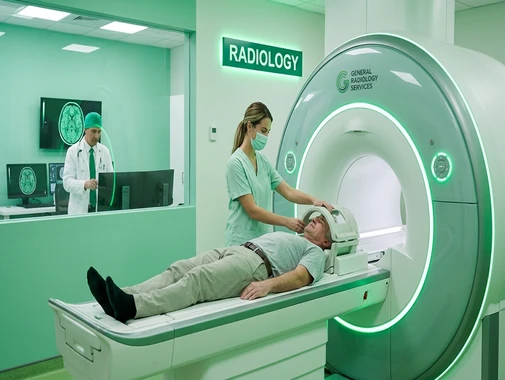
Advanced Diagnostic Imaging & Radiology Services
The Radiology Department at AS Medical Complex provides diagnostic imaging services to support the detection, diagnosis, monitoring, and treatment planning of a wide range of medical conditions.
Radiology plays an important role across almost every area of modern healthcare. Imaging can help physicians examine bones, organs, blood vessels, soft tissues, the brain, spine, chest, abdomen, pelvis, and other parts of the body without conventional surgery.
Our Radiology Department works in coordination with medical, surgical, emergency, and inpatient departments to provide imaging according to each patient's clinical requirements.
Depending on the examination required, radiological imaging may include MRI, CT Scan, X-Ray, Ultrasound, Mammography, and other specialized imaging services. The reference Radiology service similarly includes MRI, CT, digital X-ray, ultrasound, DEXA, CBCT, fluoroscopy, mammography, and image-guided procedures.
Our Radiology Services
The Radiology Department at AS Medical Complex may provide services including:
-
Magnetic Resonance Imaging – MRI
-
Computed Tomography – CT Scan
-
Digital X-Ray
-
Chest X-Ray
-
Bone and joint X-Ray
-
Ultrasound
-
Abdominal Ultrasound
-
Pelvic Ultrasound
-
Obstetric Ultrasound
-
Small-parts Ultrasound
-
Portable / bedside Ultrasound where available
-
Mammography
-
Breast imaging
-
Bone-density assessment where available
-
DEXA / DXA Scan where available
-
Fluoroscopy where available
-
Dental and maxillofacial imaging where available
-
CBCT where available
-
Contrast-enhanced imaging
-
Emergency diagnostic imaging
-
Inpatient imaging
-
Pre-operative imaging
-
Follow-up imaging
-
Image-guided procedures where available
-
Image-guided biopsy where clinically indicated
-
Radiological reporting and specialist interpretation
The appropriate imaging examination is selected according to the patient's symptoms, clinical findings, medical history, and referring physician's request.
MRI – Magnetic Resonance Imaging
Magnetic Resonance Imaging (MRI) produces detailed images of internal organs and tissues using magnetic fields and radiofrequency energy rather than ionizing X-rays.
MRI is particularly useful for evaluating soft tissues and may be requested for conditions involving:
-
Brain
-
Spine
-
Joints
-
Muscles
-
Ligaments
-
Tendons
-
Pelvic organs
-
Abdomen
-
Selected blood vessels
-
Other soft-tissue structures
The Ali Medical Radiology service similarly describes MRI as being used for neurological, musculoskeletal, and body imaging.
MRI of the Brain
Brain MRI may be requested when physicians need detailed imaging for conditions such as:
-
Persistent neurological symptoms
-
Stroke-related assessment
-
Seizures
-
Multiple sclerosis
-
Brain tumors or masses
-
Unexplained headaches with concerning features
-
Pituitary abnormalities
-
Certain infections
-
Other neurological disorders
Whether MRI is appropriate depends on the patient's symptoms and clinical assessment.
MRI of the Spine
MRI can provide detailed imaging of the spinal cord, vertebral structures, discs, nerves, and surrounding soft tissues.
It may be requested for:
-
Persistent back or neck pain with neurological symptoms
-
Disc-related conditions
-
Nerve compression
-
Spinal stenosis
-
Spinal cord abnormalities
-
Tumors
-
Infection
-
Trauma
-
Other spinal conditions
Musculoskeletal MRI
MRI can help evaluate joints and soft tissues such as:
-
Knee
-
Shoulder
-
Hip
-
Ankle
-
Wrist
-
Elbow
It may provide information about ligaments, tendons, cartilage, muscles, bone marrow, and other structures that may not be fully assessed on routine X-rays.
MRI Safety
Because MRI uses a strong magnetic field, patients should inform the radiology team before the examination if they have any implanted or metallic device.
Important information may include:
-
Pacemaker or cardiac device
-
Cochlear implant
-
Previous vascular clips
-
Metallic implants
-
Artificial joints
-
Previous surgery involving metal
-
Insulin pumps or other medical devices
-
Metal fragments from previous injury
-
Pregnancy or possible pregnancy
-
Kidney disease if MRI contrast is being considered
-
Previous reaction to contrast material
-
Claustrophobia
The radiology team determines whether a device is compatible with MRI and whether additional precautions are required.
CT Scan – Computed Tomography
Computed Tomography (CT) uses X-rays and computer processing to create detailed cross-sectional images of the body.
CT is particularly valuable when rapid and detailed imaging is required and may be used to evaluate the:
-
Brain
-
Chest
-
Abdomen
-
Pelvis
-
Bones
-
Spine
-
Blood vessels
-
Internal injuries
-
Other body regions
The reference Radiology department describes CT as providing rapid cross-sectional imaging for conditions involving the chest, abdomen, pelvis, bones, internal injury, tumors, and vascular disease.
CT Head & Brain
CT of the head may be requested in situations such as:
-
Head injury
-
Suspected intracranial bleeding
-
Acute neurological symptoms
-
Selected stroke assessment
-
Severe or sudden neurological change
-
Skull injury
-
Certain brain abnormalities
In emergency situations, CT can provide rapid imaging that helps the treating medical team make timely decisions.
CT Chest
CT imaging of the chest can provide detailed information about:
-
Lungs
-
Airways
-
Pleura
-
Mediastinum
-
Chest wall
-
Selected blood vessels
-
Other chest structures
It may be requested for:
-
Abnormal chest X-Ray
-
Lung nodules
-
Persistent respiratory symptoms
-
Chest infection complications
-
Suspected masses
-
Trauma
-
Other pulmonary or thoracic conditions
CT Abdomen & Pelvis
CT of the abdomen or pelvis can help evaluate structures including:
-
Liver
-
Gallbladder
-
Pancreas
-
Kidneys
-
Spleen
-
Bowel
-
Bladder
-
Reproductive organs
-
Lymph nodes
-
Blood vessels
Possible reasons for imaging may include:
-
Severe or persistent abdominal pain
-
Trauma
-
Infection
-
Suspected stones
-
Masses
-
Bowel disorders
-
Other abdominal or pelvic abnormalities
Contrast-Enhanced CT
Some CT examinations use contrast material to make specific organs, blood vessels, or abnormalities easier to visualize.
Before receiving contrast, patients should inform the medical team about:
-
Previous contrast reactions
-
Kidney disease
-
Diabetes medications where relevant
-
Pregnancy
-
Allergies
-
Other significant medical conditions
The radiologist or treating physician determines whether contrast is necessary.
X-Ray
X-Ray or radiography uses a small dose of ionizing radiation to create images of internal structures. It is one of the most commonly used forms of diagnostic imaging.
X-Ray may be used to evaluate:
-
Bone fractures
-
Joint injuries
-
Chest conditions
-
Lung infections
-
Certain abdominal conditions
-
Bone abnormalities
-
Foreign bodies
-
Other medical conditions
The Ali Medical Radiology department likewise identifies digital X-Ray as an important imaging tool for bones, chest conditions, fractures, infections, and other diagnostic needs.
Chest X-Ray
Chest X-Ray provides images of structures within the chest and may help physicians assess:
-
Lungs
-
Heart size
-
Chest structures
-
Pneumonia
-
Pleural fluid
-
Selected respiratory conditions
-
Certain cardiac findings
-
Other abnormalities
Depending on the findings, additional imaging such as CT may sometimes be recommended.
Bone & Joint X-Ray
X-Ray is commonly used to investigate:
-
Suspected fractures
-
Dislocations
-
Joint abnormalities
-
Arthritis-related changes
-
Bone infections
-
Certain bone lesions
-
Follow-up after orthopedic treatment
Radiological findings are interpreted together with the patient's history and clinical examination.
Ultrasound
Ultrasound uses high-frequency sound waves to create images of structures inside the body and does not use ionizing radiation.
The reference Radiology service describes ultrasound as a non-invasive imaging technique used for abdominal, pelvic, obstetric, and small-parts imaging.
Ultrasound examinations may include:
-
Abdominal Ultrasound
-
Pelvic Ultrasound
-
Obstetric Ultrasound
-
Kidney and urinary-tract Ultrasound
-
Thyroid Ultrasound
-
Breast Ultrasound
-
Scrotal Ultrasound
-
Soft-tissue Ultrasound
-
Small-parts Ultrasound
-
Selected vascular studies
-
Other examinations based on clinical need
Abdominal Ultrasound
Abdominal ultrasound may help assess organs including:
-
Liver
-
Gallbladder
-
Kidneys
-
Pancreas
-
Spleen
-
Selected abdominal blood vessels
It may be requested for symptoms or conditions such as:
-
Abdominal pain
-
Gallstones
-
Liver abnormalities
-
Kidney problems
-
Abnormal liver-function tests
-
Abdominal swelling
-
Other suspected abdominal conditions
Pelvic Ultrasound
Pelvic ultrasound can provide images of structures within the pelvis and may be used to assess:
-
Uterus
-
Ovaries
-
Bladder
-
Other pelvic structures
Depending on the patient's symptoms, it may assist in investigating:
-
Pelvic pain
-
Abnormal bleeding
-
Ovarian cysts
-
Fibroids
-
Other gynecological concerns
Obstetric Ultrasound
Ultrasound plays an important role during pregnancy and can be used at different stages depending on clinical requirements.
Obstetric ultrasound may help assess:
-
Pregnancy location
-
Fetal development
-
Gestational age
-
Fetal heartbeat
-
Number of fetuses
-
Placental location
-
Fetal growth
-
Selected anatomical features
-
Other pregnancy-related concerns
The type and timing of an obstetric scan are determined according to the pregnancy and treating obstetrician's recommendations.
Portable & Bedside Ultrasound
Portable ultrasound allows imaging to be performed at or near a patient's bedside where appropriate.
This can be particularly useful for:
-
Critically ill patients
-
Patients in intensive care
-
Patients with restricted mobility
-
Selected emergency assessments
-
Inpatients who cannot easily be transported
The Ali Medical Radiology service similarly describes portable ultrasound as useful for critically ill or immobile patients.
Mammography
Mammography is specialized breast imaging that uses low-dose X-rays to examine breast tissue.
It may be used for both screening and diagnostic assessment. Mammography plays an important role in identifying breast abnormalities and can detect some breast cancers before symptoms become noticeable.
Mammography may be requested for:
-
Routine breast screening according to appropriate guidelines
-
Breast lump assessment
-
Breast changes
-
Abnormal previous imaging
-
Follow-up of selected breast abnormalities
-
Other clinically indicated breast assessment
Patients should tell the radiology team if there is any possibility of pregnancy before mammography or other X-ray-based imaging.
Diagnostic Breast Imaging
Patients with breast symptoms may require imaging based on their age, symptoms, examination, and medical history.
Possible investigations may include:
-
Mammography
-
Breast Ultrasound
-
Additional specialized breast imaging
-
Image-guided biopsy where clinically indicated
Imaging findings are interpreted together with clinical breast assessment.
DEXA / DXA Bone Density Scan
Dual-Energy X-Ray Absorptiometry (DEXA or DXA) is an imaging test used to measure bone mineral density.
It is commonly used to assess bone loss, diagnose osteoporosis, and estimate the risk of fractures.
A bone-density scan may be recommended for selected patients with:
-
Osteoporosis risk
-
Previous fragility fracture
-
Long-term corticosteroid use
-
Medical conditions associated with bone loss
-
Other risk factors identified by their physician
The examination usually focuses on areas such as the hip and lumbar spine.
Fluoroscopy
Fluoroscopy uses X-rays to create real-time moving images.
It allows physicians to observe movement within the body or guide selected diagnostic and therapeutic procedures.
The Ali Medical Radiology service identifies fluoroscopy for dynamic examinations including gastrointestinal studies and selected image-guided procedures.
Depending on available services, fluoroscopy may be used for:
-
Gastrointestinal contrast studies
-
Swallowing-related imaging
-
Selected urinary investigations
-
Image-guided procedures
-
Other dynamic radiological examinations
The exact procedure depends on the referring physician's request and clinical indication.
CBCT – Cone Beam CT
Cone Beam Computed Tomography (CBCT) produces three-dimensional images and is particularly useful for selected dental, maxillofacial, orthodontic, and ENT applications.
The reference Radiology department includes CBCT for detailed three-dimensional imaging of dental and maxillofacial structures.
Where available, CBCT may support assessment involving:
-
Teeth and jaws
-
Dental implants
-
Impacted teeth
-
Maxillofacial structures
-
Orthodontic planning
-
Selected ENT conditions
Image-Guided Biopsy
Imaging can be used to guide a biopsy needle accurately toward a suspicious area.
Depending on the lesion and available services, guidance may involve:
-
Ultrasound
-
CT
-
Mammography
-
MRI
Image-guided biopsy allows a tissue sample to be obtained for laboratory examination without necessarily requiring open surgery. RadiologyInfo notes that image-guided biopsy can use ultrasound, MRI, or mammographic guidance depending on the abnormality being assessed.
The biopsy sample is subsequently examined by pathology to help establish a diagnosis.
Role of the Radiologist
A radiologist is a physician specializing in medical imaging.
The radiologist's role may include:
-
Reviewing imaging examinations
-
Identifying abnormalities
-
Comparing current and previous scans
-
Preparing radiology reports
-
Advising referring physicians on imaging findings
-
Recommending additional imaging when appropriate
-
Performing selected image-guided procedures
-
Participating in multidisciplinary patient care
Radiological interpretation should always be considered together with the patient's symptoms, laboratory results, medical history, and clinical examination.
Radiology Reports
After an imaging study is completed, the images are reviewed by a radiologist.
A radiology report generally communicates:
-
Type of examination
-
Relevant imaging findings
-
Important abnormalities
-
Comparison with previous studies where available
-
Radiologist's impression or conclusion
-
Recommendations for additional assessment when appropriate
Previous imaging can be particularly helpful because comparing old and new examinations may show whether an abnormality is stable, improving, or changing over time.
Importance of Previous Imaging
Patients are encouraged to bring previous:
-
X-Rays
-
CT scans
-
MRI scans
-
Ultrasound reports
-
Mammography studies
-
Radiology reports
-
Relevant medical records
Previous imaging can assist the radiologist in comparison and may help avoid unnecessary repeat examinations.
Contrast Media
Some radiology examinations require contrast material to improve visualization of particular tissues, organs, or blood vessels.
Before receiving contrast, patients should inform staff about:
-
Previous contrast reactions
-
Allergies
-
Kidney disease
-
Pregnancy
-
Current medications
-
Significant medical conditions
The use of contrast is determined according to the type of examination and individual patient's clinical situation.
Radiology & Pregnancy
Patients who are pregnant or believe they may be pregnant should tell the doctor and radiology staff before undergoing an examination involving ionizing radiation.
The radiology team can then assess:
-
Whether the examination is necessary
-
Whether another imaging method may be appropriate
-
Whether the examination can be modified
-
What precautions should be taken
Ultrasound and MRI do not use ionizing X-rays, although the choice of imaging must still be based on the patient's individual medical situation.
Radiation Safety
X-Ray, CT, mammography, fluoroscopy, and DEXA involve ionizing radiation, although the amount and purpose vary significantly between examinations.
Modern radiological practice aims to use an appropriate radiation dose while obtaining images of sufficient diagnostic quality. RadiologyInfo similarly notes the importance of minimizing radiation exposure while maintaining adequate imaging quality.
The potential benefit of medically necessary imaging is considered alongside potential radiation exposure.
Pediatric Radiology Considerations
Children can require imaging for many medical conditions, including:
-
Injuries
-
Respiratory problems
-
Abdominal symptoms
-
Neurological conditions
-
Developmental concerns
-
Congenital abnormalities
-
Other pediatric illnesses
Because children may be more sensitive to radiation, imaging techniques and examination protocols should be selected according to age, body size, and clinical need.
Ultrasound or MRI may be preferred in selected situations when these modalities can provide the required diagnostic information.
Emergency Radiology
Diagnostic imaging plays an important role in emergency medical assessment.
Depending on the patient's symptoms and condition, emergency imaging may include:
-
X-Ray
-
CT Scan
-
Ultrasound
-
MRI when appropriate and available
Imaging may help evaluate conditions such as:
-
Trauma
-
Fractures
-
Internal injuries
-
Acute neurological symptoms
-
Severe abdominal pain
-
Chest conditions
-
Kidney stones
-
Other urgent medical problems
The type of investigation is selected by the treating medical team according to the patient's clinical condition.
Radiology for Inpatients
Hospitalized patients may require imaging to:
-
Establish a diagnosis
-
Monitor an existing condition
-
Evaluate complications
-
Assess response to treatment
-
Plan surgery
-
Guide selected procedures
Radiology can support departments including:
-
Emergency Medicine
-
General Medicine
-
Pediatrics
-
Cardiology
-
Neurology
-
Pulmonology
-
Gastroenterology
-
Nephrology
-
Orthopedics
-
General Surgery
-
Gynecology & Obstetrics
-
Urology
-
Oncology
-
Critical Care
-
Other medical and surgical specialties
Pre-Operative Imaging
Imaging may be required before surgery to help surgeons understand the location and extent of disease or injury.
Depending on the condition, pre-operative imaging may include:
-
X-Ray
-
CT
-
MRI
-
Ultrasound
-
Mammography
-
Other specialized studies
The surgeon and radiologist determine the most appropriate imaging examination according to the planned procedure.
Follow-Up Imaging
Some patients require repeat imaging to monitor:
-
Healing after injury
-
Post-operative recovery
-
Tumor response
-
Chronic disease
-
Changes in a known abnormality
-
Treatment response
-
Disease progression
-
Other clinical conditions
Follow-up imaging should be performed according to clinical need rather than automatically repeating tests without an appropriate indication.
Preparing for a Radiology Examination
Preparation varies according to the type of examination.
Patients may be asked to:
-
Fast before certain studies
-
Drink water before selected ultrasound examinations
-
Remove jewelry or metallic objects
-
Wear appropriate clothing or a hospital gown
-
Provide information about implants
-
Provide previous imaging
-
Have kidney-function tests before certain contrast examinations
-
Follow specific medication instructions
Patients should follow the preparation instructions provided for their particular examination.
Information to Tell the Radiology Team
Before your imaging examination, inform the staff if you:
-
Are pregnant or may be pregnant
-
Have kidney disease
-
Have previously reacted to contrast material
-
Have a pacemaker or implanted device
-
Have metal implants
-
Have significant allergies
-
Have undergone previous surgery
-
Are taking medicines relevant to the planned procedure
-
Have severe claustrophobia before MRI
-
Have previous imaging of the same area
This information helps the team plan the examination safely and appropriately.
Coordinated Diagnostic Care
Radiology works closely with medical and surgical departments throughout the hospital.
Imaging findings can help clinicians:
-
Confirm or exclude possible diagnoses
-
Determine the location and extent of disease
-
Plan treatment
-
Plan surgery
-
Monitor response to treatment
-
Identify complications
-
Decide whether additional investigations are required
At AS Medical Complex, our aim is for Radiology to function as an integrated diagnostic service supporting comprehensive patient care.
Radiology FAQs
What is Radiology?
Radiology is the medical specialty that uses imaging technologies to help diagnose, monitor, and in some cases guide the treatment of medical conditions. Common imaging methods include X-Ray, CT, MRI, ultrasound, and mammography.
What is the difference between MRI and CT Scan?
MRI uses a strong magnetic field and radiofrequency energy to create detailed images and does not use ionizing X-rays. CT uses X-rays and computer processing to create detailed cross-sectional images. The most appropriate examination depends on the body part and medical condition being investigated.
Is ultrasound safe?
Ultrasound creates images using sound waves rather than ionizing radiation. It is widely used for abdominal, pelvic, obstetric, and other diagnostic examinations.
Do I need a contrast injection for every CT or MRI?
No. Many CT and MRI examinations can be performed without contrast. Contrast is used when it provides additional clinically useful information. The radiologist or referring physician determines whether it is appropriate for a particular examination.
Should I tell the radiology department if I am pregnant?
Yes. Always inform the radiology team if you are pregnant or think you may be pregnant, particularly before an examination involving X-rays or other ionizing radiation. This allows the team to determine the safest and most appropriate imaging approach.
What is a DEXA Scan?
DEXA or DXA is a low-dose X-ray examination used to measure bone mineral density. It is commonly used to evaluate osteoporosis and fracture risk.
Why should I bring my previous scans?
Previous scans allow radiologists to compare current findings with older images. Comparison can help determine whether an abnormality is new, stable, improving, or progressing and can sometimes prevent unnecessary repeat imaging.
Radiology Care at AS Medical Complex
The Radiology Department at AS Medical Complex is committed to providing accurate, timely, patient-focused diagnostic imaging that supports medical and surgical care across the hospital.
From routine X-Rays and Ultrasound to advanced MRI, CT, Mammography, and specialized imaging examinations, our goal is to provide high-quality diagnostic information that helps physicians make informed decisions regarding each patient's care.
Book or schedule your diagnostic imaging at AS Medical Complex for MRI, CT Scan, X-Ray, Ultrasound, Mammography, bone-density assessment, and other radiological investigations as advised by your treating physician.