
Dexa Scan at AS Medical Complex
Contact hospital
- Availability
Contact hospital
- Booking
WhatsApp / call
- Location
Islamabad
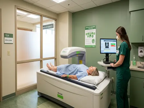
Bone Mineral Density Testing for Osteoporosis & Fracture-Risk Assessment
The DEXA Scan Service at AS Medical Complex provides Bone Mineral Density testing for patients who require assessment of bone strength, osteoporosis, low bone density, or increased risk of fragility fractures.
DEXA – more formally written as DXA, or Dual-Energy X-Ray Absorptiometry – is a specialized low-dose X-ray examination used to measure the mineral content and density of bones.
The International Society for Clinical Densitometry recommends the abbreviation DXA, although the term DEXA is also widely used by patients and healthcare providers.
DXA is most commonly used to:
-
Measure Bone Mineral Density – BMD
-
Detect low bone density
-
Diagnose osteoporosis in appropriate patients
-
Assess risk of osteoporosis-related fractures
-
Establish a baseline before selected treatment
-
Monitor osteoporosis treatment
-
Evaluate patients with medical conditions associated with bone loss
-
Assess selected patients using medicines that can weaken bone
The National Institute of Arthritis and Musculoskeletal and Skin Diseases – NIAMS – identifies DXA as the most commonly used Bone Mineral Density test and states that testing can help diagnose osteoporosis, assess fracture risk, and monitor treatment effectiveness.
At AS Medical Complex, DXA findings should be interpreted together with the patient’s:
-
Age
-
Sex
-
Menopausal status
-
Previous fractures
-
Family history
-
Medical conditions
-
Medication history
-
Body weight
-
Smoking history
-
Other fracture-risk factors
A DXA result should therefore form part of a broader bone-health assessment rather than being interpreted as an isolated number.
Our DEXA Scan Services
Depending on clinical indication, equipment capability, and available protocols, DEXA services at AS Medical Complex may include:
-
Bone Mineral Density testing
-
Osteoporosis assessment
-
Low bone-density assessment
-
Lumbar Spine DXA
-
Hip DXA
-
Femoral Neck Bone Density assessment
-
Total Hip Bone Density assessment
-
Forearm DXA where clinically indicated and available
-
Baseline bone-density testing
-
Follow-up bone-density testing
-
Osteoporosis-treatment monitoring
-
Assessment following selected fragility fractures
-
Bone-health assessment in postmenopausal women
-
Bone-health assessment in selected older adults
-
Bone-density assessment in selected men at increased risk
-
Assessment for medication-related bone loss
-
Corticosteroid-related bone-loss assessment
-
Bone-density assessment in selected endocrine disorders
-
Bone-health assessment in selected kidney disease
-
Bone-health assessment in selected liver disease
-
Bone-health assessment before selected orthopedic or spine procedures
-
Vertebral Fracture Assessment – VFA where equipment capability and clinical indication allow
-
Body-composition DXA where specifically available
-
Specialist interpretation and follow-up referral
Services such as VFA, whole-body composition analysis, pediatric DXA, or specialized forearm scanning should only be advertised as routine onsite services after confirming the exact capabilities of the AS Medical Complex DXA system.
What Is a DEXA Scan?
A DEXA or DXA scan is a specialized type of X-ray examination designed primarily to measure Bone Mineral Density.
The test passes two low-energy X-ray beams through the area being examined.
The machine measures how much of each X-ray beam is absorbed by the bones and surrounding tissue.
Computer analysis then calculates Bone Mineral Density.
DXA most commonly measures areas where osteoporosis-related fractures frequently occur, particularly the:
-
Lumbar spine
-
Hip
-
Femoral neck
NIAMS identifies hip and spine DXA as the most reliable commonly used method for diagnosing osteoporosis and estimating fracture risk.
What Is Bone Mineral Density?
Bone Mineral Density – BMD describes the amount of mineral present within a measured area of bone.
Bones containing more mineral are generally denser and tend to be stronger.
Reduced Bone Mineral Density can increase susceptibility to fractures.
Bone strength, however, depends on more than BMD alone.
Fracture risk can also be influenced by:
-
Age
-
Previous fractures
-
Falls
-
Balance
-
Muscle strength
-
Family history
-
Medication
-
Medical conditions
-
Smoking
-
Other clinical factors
For this reason, DXA contributes to fracture-risk assessment but does not represent the entire risk calculation.
What Is Osteoporosis?
Osteoporosis is a bone disease in which Bone Mineral Density, bone mass, or bone structure deteriorates sufficiently to weaken the skeleton and increase fracture risk.
Osteoporosis is sometimes described as a silent disease because a person may have substantial bone loss without obvious symptoms.
In some patients, the first indication is a fracture occurring after relatively minor trauma.
Common osteoporosis-related fracture sites include:
-
Hip
-
Spine
-
Wrist
Osteoporosis May Have No Symptoms
People sometimes expect osteoporosis to cause:
-
Generalized bone pain
-
Obvious weakness
-
Noticeable symptoms
before a fracture occurs.
This is not necessarily the case.
Bone loss can progress silently.
This is one reason appropriate bone-density assessment is important in patients who have significant osteoporosis risk factors.
What Is Osteopenia?
Osteopenia, also called low bone mass, means Bone Mineral Density is below the expected young-adult reference range but has not reached the conventional DXA threshold used to classify osteoporosis.
Using T-score classification:
-
–1.0 or higher: generally regarded as normal Bone Mineral Density
-
Between –1.0 and –2.5: low Bone Mineral Density / osteopenia
-
–2.5 or lower: may meet the densitometric definition of osteoporosis in the appropriate patient population
Osteopenia does not automatically mean that osteoporosis will develop.
However, fracture risk should be considered together with other medical and lifestyle factors.
Osteopenia vs Osteoporosis
Osteopenia
Bone Mineral Density is below normal but not within the osteoporosis range.
Osteoporosis
Bone strength is sufficiently reduced that fracture risk is substantially increased, with diagnosis based on DXA criteria and/or other clinical circumstances depending on the patient.
Treatment decisions should not be made solely from whether the report uses the word “osteopenia” or “osteoporosis.”
The treating clinician may also consider:
-
Age
-
Previous fractures
-
Family history
-
Fall risk
-
Medication
-
Other diseases
-
Overall estimated fracture risk
What Is a T-Score?
A T-score compares a patient’s Bone Mineral Density with the reference Bone Mineral Density of a healthy young adult population.
NIAMS describes the usual interpretation as:
-
T-score of –1.0 or higher: normal
-
T-score between –1.0 and –2.5: osteopenia
-
T-score of –2.5 or lower: may indicate osteoporosis
In clinical densitometry, T-scores are particularly used for osteoporosis classification in:
-
Postmenopausal women
-
Men age 50 years and older
The International Society for Clinical Densitometry states that osteoporosis may be diagnosed in these groups when the T-score at an appropriate validated skeletal site is –2.5 or below.
Lower T-Score & Fracture Risk
In general, a lower T-score indicates lower Bone Mineral Density and greater fracture risk.
However, fracture risk should not be estimated from the T-score alone.
Other important factors may include:
-
Previous low-trauma fracture
-
Increasing age
-
Family history of hip fracture
-
Smoking
-
Corticosteroid treatment
-
Falls
-
Other medical conditions
DXA should therefore be interpreted in clinical context.
What Is a Z-Score?
A Z-score compares the patient’s Bone Mineral Density with that expected in people of a similar:
-
Age
-
Sex
-
Reference population
NIAMS states that Z-scores are generally used for:
-
Premenopausal women
-
Men younger than 50
-
Children
and that a Z-score of –2.0 or lower indicates Bone Mineral Density that is low for the expected reference group and may prompt investigation for secondary causes of bone loss.
T-Score vs Z-Score
T-Score
Compares Bone Mineral Density with a healthy young-adult reference population.
It is commonly used for osteoporosis classification in postmenopausal women and men age 50 or older.
Z-Score
Compares Bone Mineral Density with an age- and sex-related reference population.
It is particularly relevant in younger adults and children.
The correct score should therefore be interpreted according to the patient’s age and clinical circumstances.
Lumbar Spine DEXA
The lumbar spine is one of the principal regions measured during central DXA.
The International Society for Clinical Densitometry recommends measurement of the posterior-anterior lumbar spine together with the hip for routine central DXA assessment.
Lumbar spine BMD can provide important information about osteoporosis.
However, interpretation can sometimes be affected by:
-
Degenerative changes
-
Previous spinal surgery
-
Vertebral abnormalities
-
Other artifacts
The reporting professional should identify vertebrae that cannot be reliably interpreted.
Hip DEXA
The hip is another major site used for osteoporosis assessment.
DXA may measure:
-
Femoral neck
-
Total hip
Hip Bone Mineral Density is particularly relevant because hip fractures can cause substantial:
-
Pain
-
Disability
-
Loss of mobility
-
Loss of independence
The ISCD recommends spine and hip BMD measurements in routine DXA assessment where clinically appropriate.
Femoral Neck Bone Density
The femoral neck is the narrow portion of bone just below the ball of the hip joint.
It is one of the recognized anatomical sites used for DXA osteoporosis classification and fracture-risk assessment.
The report may provide a femoral-neck:
-
BMD
-
T-score
-
Z-score
depending on the patient.
Forearm DEXA
Forearm Bone Mineral Density may be measured in selected circumstances.
The ISCD recommends considering forearm measurement when:
-
Hip or spine cannot be measured or interpreted
-
Hyperparathyroidism is present
-
Patient size exceeds the DXA table limit
Forearm DXA should therefore not automatically replace standard central DXA of the hip and spine.
Availability at AS Medical Complex should be confirmed.
Who May Need a DEXA Scan?
A clinician may consider DXA for patients who have increased risk of osteoporosis or fracture.
Factors may include:
-
Increasing age
-
Menopause
-
Previous low-trauma fracture
-
Family history of osteoporosis
-
Family history of hip fracture
-
Long-term corticosteroid therapy
-
Low body weight
-
Smoking
-
Selected endocrine disorders
-
Chronic kidney disease
-
Chronic liver disease
-
Rheumatoid arthritis or other inflammatory disease
-
Significant immobility
-
Other conditions associated with bone loss
RadiologyInfo identifies many of these as circumstances in which Bone Mineral Density testing may be particularly useful.
DEXA Screening in Women
Screening recommendations vary between countries and professional organizations.
For example, the current U.S. Preventive Services Task Force recommendation advises osteoporosis screening in:
-
Women age 65 years or older
-
Postmenopausal women younger than 65 who are at increased fracture risk based on clinical risk assessment
These are U.S. recommendations and should not automatically be treated as the universal screening policy for every patient in Pakistan.
At AS Medical Complex, screening decisions should take account of:
-
Patient age
-
Menopausal status
-
Individual risk factors
-
Medical history
-
Relevant locally applicable guidance
DEXA Screening in Men
Men can also develop osteoporosis.
Risk may increase with:
-
Older age
-
Low body weight
-
Previous fracture
-
Chronic illness
-
Long-term corticosteroid use
-
Hypogonadism
-
Other medical conditions
NIAMS confirms that osteoporosis is an important condition in men and that central DXA is the most commonly used test for diagnosis.
Population-screening recommendations for men differ between professional organizations.
For example, the 2025 USPSTF concluded that evidence was insufficient to determine the balance of benefits and harms of routine population osteoporosis screening in men.
This does not mean that men with clinical risk factors should never undergo DXA.
Individual assessment remains important.
Postmenopausal Women
Loss of estrogen after menopause can accelerate bone loss.
Postmenopausal women may therefore have increased risk of:
-
Osteopenia
-
Osteoporosis
-
Fragility fractures
DXA may be recommended according to age and individual fracture-risk factors.
Early Menopause
Women who experience menopause at a younger age may have a longer period of reduced estrogen exposure.
A clinician may consider their:
-
Age at menopause
-
Other medical conditions
-
Previous fractures
-
Body weight
-
Medication
-
Additional risk factors
when deciding whether Bone Mineral Density testing is appropriate.
Long-Term Corticosteroid Use
Medicines such as glucocorticoids can contribute to bone loss when used for prolonged periods.
Examples include patients using corticosteroids for selected:
-
Rheumatological diseases
-
Autoimmune disorders
-
Respiratory diseases
-
Other chronic conditions
RadiologyInfo and ISCD identify chronic corticosteroid exposure as an important osteoporosis and DXA consideration.
Patients should not stop prescribed steroids suddenly because of concern about bone health.
Bone protection and medication decisions should be discussed with the treating doctor.
Rheumatoid Arthritis & Bone Health
Inflammatory diseases such as rheumatoid arthritis may increase fracture risk through several mechanisms, including:
-
Chronic inflammation
-
Reduced mobility
-
Corticosteroid exposure
-
Other factors
Bone-health assessment may therefore be appropriate in selected patients.
The ISCD includes inflammatory arthritis among conditions associated with impaired bone health in selected orthopedic and surgical populations.
Kidney Disease & Bone Density
Chronic Kidney Disease can affect:
-
Bone metabolism
-
Calcium
-
Phosphorus
-
Vitamin D
-
Parathyroid hormone
-
Bone strength
Patients with significant kidney disease may have bone disorders that are more complex than ordinary postmenopausal osteoporosis.
The ISCD identifies advanced Chronic Kidney Disease as an important risk factor for impaired bone health in selected patients.
Bone-density results should therefore be interpreted with the Nephrology team where appropriate.
Hyperparathyroidism
Parathyroid disease can alter calcium metabolism and reduce Bone Mineral Density.
RadiologyInfo notes that DXA may be used to assess bone complications of parathyroid disease.
Patients with suspected hyperparathyroidism may also require:
-
Calcium testing
-
Parathyroid hormone testing
-
Vitamin D testing
-
Kidney assessment
-
Endocrinology consultation
Thyroid Disease
Certain thyroid conditions or excessive thyroid-hormone exposure can contribute to bone loss.
Patients with thyroid disease may require assessment of:
-
TSH
-
Thyroid hormones
-
Medication dose
-
Other bone-health risks
before deciding whether DXA is appropriate.
Low Body Weight
Low body weight can be associated with increased osteoporosis and fracture risk.
Assessment may consider:
-
Nutrition
-
Menstrual history
-
Chronic disease
-
Gastrointestinal disease
-
Eating disorders
-
Other causes of low weight
A DEXA scan may be appropriate when low body weight forms part of a wider osteoporosis-risk profile.
Smoking & Bone Health
Smoking is associated with increased osteoporosis and fracture risk.
NIAMS includes smoking among recognized osteoporosis risk factors.
Smoking cessation can therefore support both:
-
Bone health
-
General cardiovascular and respiratory health
Previous Fragility Fracture
A fragility fracture is a fracture occurring after relatively minor trauma that would not normally break healthy bone.
Examples may include a fracture after:
-
Falling from standing height
-
Minor impact
-
Other low-energy trauma
A previous fragility fracture is an important sign that bone strength may be impaired and can substantially affect future fracture risk.
Such patients may require:
-
Bone-density assessment
-
Medical evaluation for osteoporosis
-
Treatment
-
Fall-risk assessment
Hip Fracture & Osteoporosis
Hip fractures in older adults are commonly associated with osteoporosis.
They can lead to:
-
Surgery
-
Hospitalization
-
Reduced mobility
-
Rehabilitation
-
Loss of independence
-
Increased risk of another fracture
Patients with an osteoporosis-related fracture should receive broader bone-health assessment rather than treatment of the broken bone alone.
Vertebral Compression Fractures
Osteoporosis can cause vertebral compression fractures in the spine.
These may result in:
-
Back pain
-
Loss of height
-
Stooped posture
-
Reduced mobility
Some vertebral fractures may occur with relatively little trauma.
DXA measures Bone Mineral Density but is not itself a complete diagnostic test for every acute spinal fracture.
A patient with new severe back pain may require:
-
X-Ray
-
CT
-
MRI
-
Other assessment
depending on symptoms.
Vertebral Fracture Assessment – VFA
Some DXA systems can perform Vertebral Fracture Assessment – VFA.
This is a low-dose lateral imaging examination used to look for selected vertebral fractures.
RadiologyInfo notes that VFA can be performed on some DXA systems and generally adds only a short additional period to the examination.
The ISCD recommends VFA in selected patients based on clinical risk factors.
VFA availability at AS Medical Complex should be confirmed before it is promoted as a routine service.
Loss of Height
Significant loss of height can sometimes suggest vertebral compression fractures.
Patients who notice:
-
Progressive height loss
-
Increasing stooped posture
-
New back pain
may require clinical assessment.
The doctor may consider:
-
DXA
-
VFA
-
Spine X-Ray
-
MRI
-
Other investigations
depending on the presentation.
DEXA Before Orthopedic or Spine Surgery
Bone quality can influence the planning and outcome of some orthopedic and spine procedures.
The ISCD recommends considering bone-health assessment in patients undergoing elective orthopedic or spine surgery when they meet appropriate DXA testing criteria.
Selected higher-risk patients can include those with:
-
Long-standing diabetes
-
Inflammatory arthritis
-
Chronic corticosteroid exposure
-
Low-trauma fracture after age 50
-
Advanced Chronic Kidney Disease
-
Limited mobility
-
Smoking history
The Orthopedic or Spine Surgery team should determine when DXA is appropriate.
DEXA & Diabetes
Diabetes can affect fracture risk in ways that are not always fully represented by Bone Mineral Density alone.
Patients with long-standing diabetes may therefore require broader bone-health and fracture-risk assessment.
The ISCD identifies long-duration, poorly controlled diabetes as a risk factor for impaired bone health in selected orthopedic patients.
DEXA & Cancer Treatment
Some cancer treatments can reduce bone density.
Examples may include therapies that significantly reduce:
-
Estrogen
-
Testosterone
-
Other hormonal influences on bone
Selected cancer patients may therefore require Bone Mineral Density monitoring according to their Oncology treatment plan.
The exact timing should be determined by the treating team.
DEXA & Bariatric or Major Weight-Loss Treatment
Significant weight loss and some bariatric procedures can affect:
-
Bone density
-
Nutrition
-
Vitamin D
-
Calcium metabolism
-
Muscle mass
Specialized DXA body-composition or Bone Mineral Density assessment may be useful in selected patients.
The ISCD recognizes total-body DXA body-composition analysis in certain patients undergoing substantial weight loss, though its clinical role depends on the individual situation.
Availability of body-composition DXA should be confirmed at AS Medical Complex.
DEXA Is Not the Same as a Regular X-Ray
A standard X-ray can show:
-
Fractures
-
Bone alignment
-
Certain obvious bone abnormalities
but is not sensitive enough to precisely measure Bone Mineral Density.
DXA is specifically designed to quantify bone density.
A person can have osteoporosis even when an ordinary X-ray does not clearly show severe bone loss.
DEXA vs CT Scan
CT can provide detailed anatomical images and, in selected settings, specialized quantitative CT can estimate bone density.
However, DXA remains the established standard examination for routine Bone Mineral Density measurement.
The ISCD generally prefers central DXA at the spine and femur for therapeutic decisions when it can be performed appropriately.
DEXA vs MRI
MRI provides highly detailed information about:
-
Bone marrow
-
Soft tissues
-
Spinal cord
-
Muscles
-
Ligaments
-
Other structures
but routine MRI is not used as the standard test for osteoporosis Bone Mineral Density classification.
DXA and MRI therefore answer different clinical questions.
DEXA vs Bone Scan
A DEXA scan should not be confused with a nuclear medicine bone scan.
DEXA
Measures Bone Mineral Density.
Nuclear Medicine Bone Scan
Uses a radioactive tracer to identify selected areas of abnormal bone activity.
The investigations have different purposes and should not be considered interchangeable.
DEXA vs Peripheral Bone Screening
Some portable or peripheral devices measure bone density at sites such as:
-
Wrist
-
Heel
-
Forearm
NIAMS notes that peripheral DXA can help identify low bone mass, but it may not provide the same information for predicting fractures or monitoring osteoporosis medication as central DXA of the spine and hip.
When available, central DXA is therefore preferred for many diagnostic and treatment decisions.
Fracture-Risk Assessment
Bone density is an important part of fracture-risk assessment, but it is not the only factor.
Doctors may consider:
-
Age
-
Sex
-
Weight
-
Previous fractures
-
Family history
-
Smoking
-
Alcohol
-
Corticosteroid treatment
-
Medical conditions
-
Bone Mineral Density
RadiologyInfo specifically notes that age, body weight, prior fractures, family history, smoking and alcohol consumption can affect fracture risk and treatment decisions.
DEXA Does Not Predict Exactly Who Will Break a Bone
A patient with osteoporosis does not necessarily experience a fracture.
Similarly, fractures can occur in people who do not meet the densitometric osteoporosis threshold.
DXA helps estimate risk rather than predicting with certainty whether an individual fracture will occur.
Clinical risk assessment therefore remains important.
Preparing for a DEXA Scan
DEXA requires relatively little preparation.
RadiologyInfo advises patients to:
-
Wear comfortable clothing
-
Avoid metal items where possible
-
Inform staff if pregnancy is possible
-
Mention recent barium examinations
-
Mention recent CT contrast
-
Mention recent radioisotope examinations
The AS Medical Complex imaging team should provide any examination-specific instructions.
Calcium Supplements Before DEXA
RadiologyInfo recommends avoiding calcium supplements for at least 24 hours before a DXA examination.
Patients should distinguish between:
-
Calcium supplements
-
Normal food intake
-
Other medications
and should follow the specific instructions provided by the imaging department.
Do not stop important prescription medicines unless a healthcare professional advises you to do so.
Fasting Before DEXA
Routine DXA generally does not require fasting.
Patients can usually maintain their usual eating pattern unless another test or medical procedure scheduled on the same day requires fasting.
The imaging department should provide any specific preparation instructions.
Clothing for DEXA
Loose, comfortable clothing is generally appropriate.
Patients may be asked to remove objects containing metal, such as:
-
Belt buckles
-
Metal buttons
-
Zippers
-
Keys
-
Jewelry
because these can interfere with imaging.
A hospital gown may be provided where necessary.
Recent Barium or Contrast Examinations
Patients should tell the DXA team if they have recently undergone:
-
Barium examination
-
Contrast-enhanced CT
-
Radioisotope or nuclear medicine examination
RadiologyInfo notes that these examinations may affect DXA scheduling and that the scan may sometimes need to be delayed.
The Radiology team should determine appropriate timing.
Pregnancy & DEXA
Patients should tell the healthcare team if:
-
They are pregnant
-
They think they may be pregnant
DXA uses a very small amount of ionizing radiation, but non-urgent X-ray-based examinations are generally avoided during pregnancy unless there is a clear medical reason.
RadiologyInfo specifically advises patients to inform the doctor and technologist if pregnancy is possible before DXA.
What Happens During a DEXA Scan?
During a typical central DXA examination:
-
Patient identity is confirmed.
-
Relevant clinical information is reviewed.
-
The patient lies on a padded examination table.
-
The body is positioned for the required measurement.
-
A scanning arm moves over the body.
-
Low-dose X-rays are used to measure Bone Mineral Density.
-
Measurements are processed by computer.
The examination is:
-
Non-invasive
-
Painless
-
Usually relatively quick
Positioning for Lumbar Spine DEXA
For lumbar-spine measurement, the patient may have the legs positioned or supported in a way that helps flatten the lower spine appropriately for scanning.
Correct positioning is important because inconsistent positioning can affect measurement accuracy.
Positioning for Hip DEXA
For hip DXA, the leg is positioned carefully so that the proximal femur can be measured consistently.
Small differences in rotation or positioning can influence the measurement.
Standardized technique is therefore important, particularly for follow-up examinations.
Is DEXA Painful?
DXA itself is painless.
No needle or injection is required for routine Bone Mineral Density scanning.
No anesthesia is usually required.
Patients with severe back or hip pain may feel some temporary discomfort simply because they need to remain in the required position.
They should tell the technologist if positioning is difficult.
How Long Does a DEXA Scan Take?
RadiologyInfo notes that a Bone Density examination is often completed in approximately 10 to 30 minutes, depending on the equipment and body areas examined.
AS Medical Complex should provide an estimated duration according to the specific examination rather than guaranteeing one fixed time for every patient.
Radiation Dose From DEXA
DEXA uses a very small amount of ionizing radiation.
RadiologyInfo’s current radiation-dose comparison estimates an effective dose of approximately 0.001 mSv for Bone Densitometry, comparable to only a few hours of normal background radiation.
The precise dose can vary according to:
-
Equipment
-
Protocol
-
Body area
-
Patient factors
but DXA is generally regarded as a very low-dose X-ray examination.
Does Radiation Stay in the Body?
No.
No radiation remains inside the body after a standard DXA X-ray examination.
The patient does not become radioactive after the scan.
After a DEXA Scan
Routine DXA generally requires no recovery period.
Most patients can return to their normal activities immediately after the examination.
There is usually:
-
No anesthesia
-
No injection
-
No sedation
-
No special recovery
unless another procedure has been performed at the same visit.
DEXA Results
A DXA report may include:
-
Bone Mineral Density – BMD
-
T-score
-
Z-score
-
Anatomical sites measured
-
Comparison with previous examinations where appropriate
-
Other technical or clinical information
The report should be interpreted according to:
-
Patient age
-
Sex
-
Menopausal status
-
Medical history
-
Fracture history
-
Medication
-
Other risk factors
Normal DEXA Does Not Mean Zero Fracture Risk
A normal Bone Mineral Density result is reassuring but does not guarantee that a person can never experience a fracture.
Fracture risk can also be affected by:
-
Falls
-
Trauma
-
Balance
-
Muscle weakness
-
Vision
-
Other health conditions
Bone health and fall prevention should therefore be considered together.
Low DEXA Result Does Not Automatically Mean Medication
Not every patient with osteopenia automatically requires prescription osteoporosis medication.
Treatment decisions depend on:
-
Degree of bone loss
-
Age
-
Fracture history
-
Overall fracture risk
-
Medical conditions
-
Medication history
The treating clinician should determine whether management involves:
-
Lifestyle changes
-
Nutrition
-
Exercise
-
Supplements where appropriate
-
Prescription treatment
-
Monitoring
Osteoporosis Treatment
Treatment for osteoporosis aims primarily to reduce fracture risk.
Depending on the patient, management may include:
-
Weight-bearing activity
-
Muscle-strengthening exercise
-
Balance and fall-prevention strategies
-
Appropriate calcium intake
-
Appropriate vitamin D
-
Smoking cessation
-
Medication when clinically indicated
-
Treatment of underlying secondary causes
NIAMS identifies appropriate exercise, nutrition, calcium/vitamin D, and prescription therapy among important components of osteoporosis management.
Exercise & Bone Health
Weight-bearing and muscle-strengthening activity can support bone health and reduce fall risk in appropriately selected patients.
NIAMS recommends a combination of:
-
Weight-bearing exercise
-
Resistance or strengthening exercise
-
Balance-related activity
according to the person’s health and physical capability.
Patients with established osteoporosis or previous fractures should obtain professional advice before beginning high-impact or strenuous exercise.
Calcium & Bone Health
Calcium is important for:
-
Bone structure
-
Muscle function
-
Nerve function
-
Other body processes
Inadequate calcium intake over time can contribute to reduced bone strength.
NIAMS notes that when dietary calcium is insufficient, the body may draw calcium from the bones.
Calcium supplementation should be individualized rather than automatically taken in very high doses.
Vitamin D & Bone Health
Vitamin D supports calcium absorption and bone metabolism.
Some patients at risk of deficiency may require:
-
Dietary assessment
-
Vitamin D testing
-
Supplementation
depending on the clinical circumstances.
Supplement doses should be guided by medical or nutritional assessment when possible.
Fall Prevention
Preventing falls can be as important as improving Bone Mineral Density.
Fall-risk reduction may involve:
-
Strength and balance training
-
Reviewing vision
-
Reviewing medications
-
Appropriate footwear
-
Removing household hazards
-
Using walking aids when necessary
-
Physiotherapy
A fracture usually results from a combination of bone strength and the force placed on the bone.
Monitoring Osteoporosis Treatment
DXA can be used to monitor response to osteoporosis therapy.
However, repeat testing should have a clear clinical purpose.
The ISCD recommends follow-up BMD testing when the result is expected to influence patient management and notes that intervals should be individualized according to clinical circumstances.
When Should DEXA Be Repeated?
There is no single repeat interval appropriate for every patient.
The timing can depend on:
-
Baseline BMD
-
Age
-
Treatment
-
New fracture
-
New risk factors
-
Medication changes
-
Rate of expected bone loss
-
Clinical management plan
The ISCD recommends individualizing repeat BMD testing and performing it when results are likely to influence treatment decisions.
Same Machine for Follow-Up DEXA
Comparing Bone Mineral Density over time requires careful quality control.
The ISCD notes that measurements from different DXA devices cannot always be directly compared unless appropriate cross-calibration has been performed.
For monitoring purposes, patients may therefore benefit from having follow-up scans:
-
At the same facility
-
On the same appropriately maintained device
where practical.
Small Changes May Not Be Significant
A small difference between two DXA results does not automatically mean that the patient has truly gained or lost bone.
Every DXA facility has measurement precision limits.
The ISCD recommends facilities calculate their own precision error and Least Significant Change – LSC when interpreting serial measurements.
The treating clinician should therefore interpret changes rather than simply comparing two numbers.
DEXA & Endocrinology
The Endocrinology Department may become involved when low Bone Mineral Density is associated with conditions such as:
-
Hyperparathyroidism
-
Hyperthyroidism
-
Hypogonadism
-
Other hormonal disorders
-
Complex osteoporosis
The Endocrinologist may investigate whether bone loss has a secondary medical cause.
DEXA & Rheumatology
Rheumatology may coordinate bone-health management in selected patients with:
-
Rheumatoid arthritis
-
Chronic inflammatory disease
-
Long-term corticosteroid treatment
-
Osteoporosis
-
Previous fragility fracture
Treatment may require management of both the underlying inflammatory condition and bone health.
DEXA & Orthopedics
Orthopedics may encounter osteoporosis during treatment of:
-
Hip fracture
-
Wrist fracture
-
Vertebral fracture
-
Other fragility fractures
A fracture after relatively minor trauma can be an opportunity to investigate underlying bone weakness.
The goal should not only be to heal the current fracture but also to reduce the chance of another fracture.
DEXA & Ortho & Spine Surgery
Selected spine-surgery patients may require preoperative bone-health assessment.
The ISCD recommends DXA evaluation in higher-risk elective orthopedic and spine patients who meet appropriate indications.
Bone quality can be relevant when planning:
-
Spinal fixation
-
Fusion
-
Other orthopedic procedures
DEXA & Nephrology
Patients with Chronic Kidney Disease can have complex disorders of bone and mineral metabolism.
Nephrology may need to interpret DXA alongside:
-
Calcium
-
Phosphorus
-
Vitamin D
-
Parathyroid hormone
-
Kidney function
-
Other laboratory findings
DXA alone cannot characterize every type of kidney-related bone disease.
DEXA & Gynecology / Well Women Clinic
Bone health becomes particularly important during and after menopause.
Gynecology or the Well Women Clinic may help identify women who have:
-
Early menopause
-
Other osteoporosis risk factors
-
Previous fractures
-
Long-term hormonal changes affecting bone health
and refer appropriately for Bone Mineral Density assessment.
DEXA & Dietitian Services
Dietitian support may complement osteoporosis management through assessment of:
-
Calcium intake
-
Vitamin D-related nutrition
-
Protein intake
-
Overall dietary quality
-
Low body weight
-
Malnutrition
Nutrition should form part of comprehensive bone care rather than replacing appropriate medical treatment.
DEXA & Physiotherapy
Physiotherapy can support patients with osteoporosis through individualized strategies involving:
-
Strength
-
Balance
-
Posture
-
Walking
-
Fall prevention
-
Safe physical activity
Patients with previous vertebral or other fragility fractures may need modified exercise programs to avoid inappropriate loading.
Pediatric DEXA
Children occasionally require DXA in selected medical conditions associated with impaired bone development.
However, interpretation is different from adult osteoporosis assessment.
The ISCD states that DXA is a preferred method for pediatric BMD assessment but emphasizes age-appropriate skeletal sites and interpretation.
Adult T-score diagnostic thresholds should not simply be applied to children.
Pediatric DXA should only be provided where appropriate expertise and protocols are available.
Body Composition DEXA
Some DXA systems can also measure components of body composition such as:
-
Fat mass
-
Lean mass
-
Bone mass
The ISCD recognizes specialized clinical applications for DXA body-composition analysis in selected patient groups.
Body-composition capability depends on the specific DXA machine and software installed.
It should therefore only be advertised by AS Medical Complex if confirmed.
Limitations of DEXA
DXA is a highly useful standardized test, but it has limitations.
Results may be affected by:
-
Degenerative spinal changes
-
Previous fractures
-
Surgical hardware
-
Abnormal vertebral anatomy
-
Patient positioning
-
Technical differences between machines
-
Other artifacts
It also measures Bone Mineral Density rather than every aspect of bone quality.
The final interpretation should therefore consider both imaging quality and clinical risk.
DEXA Does Not Diagnose Acute Fractures
DEXA is primarily a Bone Mineral Density test.
It should not be used as the main emergency imaging examination for a patient who has just suffered a significant injury.
A suspected acute fracture may require:
-
X-Ray
-
CT Scan
-
MRI
-
Other assessment
depending on the injury.
When Should You Consider a DEXA Scan?
You may need a DEXA scan if your doctor is assessing:
-
Osteoporosis risk
-
Low Bone Mineral Density
-
Previous fragility fracture
-
Postmenopausal bone health
-
Long-term corticosteroid use
-
Medical conditions associated with bone loss
-
Treatment response
-
Selected preoperative orthopedic bone-health concerns
-
Other clinically important osteoporosis risk factors
Screening should be individualized according to age, sex, medical history, fracture risk, and applicable clinical guidance.
When Should You See a Doctor Before Booking DEXA?
Medical assessment may be useful before DXA if you have:
-
Unexplained fractures
-
Significant loss of height
-
Persistent back pain
-
Long-term steroid use
-
Endocrine disease
-
Chronic Kidney Disease
-
Major nutritional problems
-
Other medical conditions affecting bone health
The doctor may also recommend investigations such as:
-
Calcium
-
Vitamin D
-
Kidney-function tests
-
Thyroid-function tests
-
Parathyroid hormone
-
Other laboratory testing
depending on the suspected cause.
When Is Emergency Care Required Instead of DEXA?
DEXA is generally a routine or planned investigation rather than an emergency test.
Do not wait for a DEXA appointment if you develop:
-
Severe pain after a fall
-
Obvious limb deformity
-
Inability to bear weight after injury
-
Suspected hip fracture
-
Major trauma
-
New severe back pain after trauma
-
Sudden weakness or numbness
-
New bladder or bowel dysfunction
-
Other potentially serious acute symptoms
These situations require clinical assessment and may require:
-
X-Ray
-
CT
-
MRI
-
Emergency treatment
DEXA can be considered later if underlying osteoporosis or bone weakness is suspected.
Multidisciplinary Bone-Health Care
Depending on the DEXA findings and underlying condition, care may involve:
-
Radiology
-
Endocrinology
-
Rheumatology
-
Orthopedics
-
Ortho & Spine Surgery
-
General Medicine
-
Nephrology
-
Gynecology
-
Well Women Clinic
-
Dietitian Services
-
Physiotherapy
-
Oncology
-
Other relevant specialties
The goal is not simply to identify low Bone Mineral Density but to reduce future fracture risk.
Follow-Up After a DEXA Scan
The next step depends on:
-
T-score or Z-score
-
Previous fractures
-
Age
-
Medical conditions
-
Medication
-
Overall fracture risk
Possible recommendations may include:
-
No immediate treatment
-
Lifestyle modification
-
Exercise
-
Fall prevention
-
Dietary assessment
-
Calcium or vitamin D management
-
Laboratory investigation
-
Osteoporosis medication
-
Specialist referral
-
Repeat DXA at an appropriate interval
NIAMS advises reviewing BMD results with a healthcare professional because diagnosis and treatment depend on the scan together with age and other fracture-risk factors.
Long-Term Bone-Health Monitoring
Bone health requires ongoing attention in patients with:
-
Osteoporosis
-
Significant osteopenia
-
Previous fragility fracture
-
Long-term corticosteroid therapy
-
Other chronic bone-loss risk factors
Follow-up may involve:
-
Repeat DXA where appropriate
-
Medication review
-
Calcium and vitamin D assessment
-
Exercise
-
Fall-risk assessment
-
Treatment of secondary causes
Repeat scanning should have a clear management objective rather than simply being performed at arbitrary intervals.
Dexa Scan FAQs
What is a DEXA or DXA Scan?
DEXA or DXA – Dual-Energy X-Ray Absorptiometry – is a specialized low-dose X-ray examination used to measure Bone Mineral Density. It is most commonly performed at the lumbar spine and hip and is the standard imaging method for diagnosing osteoporosis in appropriate patients, assessing fracture risk, and monitoring osteoporosis treatment.
What do T-scores of –1 and –2.5 mean?
According to NIAMS, a T-score of –1.0 or higher is generally regarded as normal Bone Mineral Density, a score between –1.0 and –2.5 represents low Bone Mineral Density or osteopenia, and a score of –2.5 or lower may indicate osteoporosis in the appropriate patient population. The result should still be interpreted with other fracture-risk factors.
Does a DEXA Scan use radiation?
Yes, but DXA uses a very small amount of ionizing radiation. RadiologyInfo lists the approximate effective radiation dose from Bone Densitometry at around 0.001 mSv, comparable to only a few hours of normal natural background radiation.
Do I need to fast before a DEXA Scan?
Routine DXA generally does not require fasting. RadiologyInfo recommends avoiding calcium supplements for at least 24 hours before the examination and informing the imaging team about recent barium studies, CT contrast, radioisotope examinations, or possible pregnancy.
Who should have a DEXA Scan?
DXA may be appropriate for people at increased risk of osteoporosis because of age, menopause, previous fragility fracture, long-term corticosteroid use, selected chronic diseases, low body weight, family history, or other risk factors. Screening guidelines vary internationally, so the decision should be individualized rather than applying one age threshold to everyone.
How often should a DEXA Scan be repeated?
There is no universal interval appropriate for every patient. The timing depends on baseline Bone Mineral Density, treatment, age, new fractures, new risk factors, and whether the result is likely to change management. The ISCD recommends individualized follow-up testing with clearly defined clinical objectives.
Can a DEXA Scan diagnose a broken bone?
DEXA is designed primarily to measure Bone Mineral Density and assess osteoporosis-related fracture risk. It is not the main diagnostic examination for an acute injury. A suspected new fracture may require X-Ray, CT Scan, MRI, or another appropriate examination. DXA can later help determine whether underlying osteoporosis contributed to a low-trauma fracture.
DEXA Scan Services at AS Medical Complex
The DEXA Scan Service at AS Medical Complex is designed to provide clinically appropriate Bone Mineral Density assessment for patients at risk of osteoporosis, osteopenia, and fragility fractures.
From lumbar-spine and hip Bone Mineral Density measurement to osteoporosis assessment, fracture-risk evaluation, baseline testing, and monitoring of treatment-related changes, our goal is to help patients and clinicians identify reduced bone strength before it results in preventable complications.
DXA is quick, non-invasive, painless, and uses a very low amount of ionizing radiation. It remains one of the most standardized and widely accepted methods for Bone Mineral Density assessment.
A DEXA result should not be considered in isolation. Appropriate bone-health care may also involve assessment of age, previous fractures, falls, medication, hormonal health, kidney disease, nutrition, calcium and vitamin D status, exercise, and other clinical risk factors.
DEXA findings can be coordinated with relevant departments at AS Medical Complex, including Radiology, Endocrinology, Rheumatology, Orthopedics, Ortho & Spine Surgery, General Medicine, Nephrology, Gynecology, Well Women Clinic, Dietitian Services, Physiotherapy, and other specialties where clinically appropriate.
Book a DEXA Scan at AS Medical Complex for physician-recommended Bone Mineral Density measurement, osteoporosis assessment, osteopenia evaluation, fracture-risk assessment, baseline testing, and clinically indicated follow-up of bone health.