
X-Ray Services at AS Medical Complex
Contact hospital
- Availability
Contact hospital
- Booking
WhatsApp / call
- Location
Islamabad
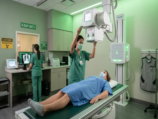
Diagnostic Radiography for Fast & Reliable Medical Imaging
The X-Ray Service at AS Medical Complex provides diagnostic radiography for patients who require imaging of the chest, bones, joints, spine, abdomen, and other clinically appropriate areas.
X-ray – also known as radiography – is one of the most widely used forms of medical imaging.
It uses a controlled amount of ionizing radiation to produce images of structures inside the body.
Different tissues absorb X-rays differently.
Dense structures such as bone absorb more X-rays and therefore appear relatively white on an X-ray image, while air appears dark and soft tissues generally appear in varying shades of gray.
X-ray imaging is particularly useful because it is:
-
Fast
-
Non-invasive
-
Widely used in emergency care
-
Useful for many bone and joint conditions
-
Useful for chest assessment
-
Helpful in evaluating selected abdominal conditions
-
Useful for postoperative assessment
-
Appropriate as an initial imaging investigation for many conditions
At AS Medical Complex, X-rays should be performed according to a valid clinical indication and interpreted alongside the patient’s symptoms, medical history, physical examination, and other investigations.
Our X-Ray Services
Depending on clinical indication and available imaging facilities, X-Ray services at AS Medical Complex may include:
-
Chest X-Ray
-
Chest PA View
-
Chest AP View where appropriate
-
Lateral Chest X-Ray where indicated
-
Portable Chest X-Ray where available
-
Skull X-Ray for selected indications
-
Facial Bone X-Ray for selected indications
-
Sinus X-Ray where clinically appropriate
-
Neck soft-tissue X-Ray where appropriate
-
Cervical Spine X-Ray
-
Thoracic Spine X-Ray
-
Lumbar Spine X-Ray
-
Lumbosacral Spine X-Ray
-
Pelvis X-Ray
-
Hip X-Ray
-
Knee X-Ray
-
Ankle X-Ray
-
Foot X-Ray
-
Shoulder X-Ray
-
Clavicle X-Ray
-
Humerus X-Ray
-
Elbow X-Ray
-
Forearm X-Ray
-
Wrist X-Ray
-
Hand X-Ray
-
Femur X-Ray
-
Tibia and Fibula X-Ray
-
Bone X-Ray
-
Joint X-Ray
-
Fracture assessment
-
Post-reduction X-Ray
-
Postoperative orthopedic X-Ray
-
Follow-up fracture X-Ray
-
Abdominal X-Ray
-
KUB X-Ray where clinically indicated
-
Selected pediatric X-Rays
-
Emergency X-Ray imaging
-
Inpatient X-Ray imaging
-
Portable radiography where available
-
Preoperative X-Ray where medically indicated
-
Follow-up radiography
-
Specialist radiologist reporting
Specific X-Ray views and examinations should be selected according to the medical question rather than applying the same views to every patient.
What Is an X-Ray?
An X-ray examination uses a controlled beam of ionizing radiation to create an image of structures inside the body.
During the examination:
-
The X-ray beam is directed toward the area being examined.
-
X-rays pass through the body.
-
Different tissues absorb different amounts of radiation.
-
A detector records the pattern of X-rays passing through the body.
-
The resulting information creates the diagnostic image.
Bone absorbs much of the X-ray beam and appears relatively white.
Soft tissues appear in different shades of gray.
Air-containing structures, such as the lungs, generally appear darker.
Digital Radiography
Modern X-ray systems may use digital detectors rather than traditional film.
Digital radiography allows images to be:
-
Displayed electronically
-
Reviewed by the radiologist
-
Enlarged for closer inspection
-
Adjusted for visualization
-
Stored electronically
-
Compared with previous examinations
-
Shared with the treating clinical team
The exact X-ray equipment installed at AS Medical Complex should be confirmed before advertising specific technical capabilities.
What Can an X-Ray Diagnose?
X-rays can help doctors investigate many different conditions.
Depending on the body area, radiography may help identify or assess:
-
Bone fractures
-
Joint dislocation
-
Bone alignment
-
Arthritis-related changes
-
Chest infection
-
Pneumonia
-
Selected lung abnormalities
-
Pleural fluid
-
Heart enlargement on chest imaging
-
Bowel-gas patterns
-
Selected bowel obstruction
-
Certain foreign bodies
-
Postoperative changes
-
Medical-device positioning in selected settings
-
Other clinically appropriate conditions
Radiography is widely used for orthopedic evaluation and many other examinations requiring a static diagnostic image.
X-Ray for Bone Fractures
One of the most common reasons for X-ray imaging is suspected fracture.
An X-ray may help determine:
-
Whether a bone is fractured
-
Location of the fracture
-
Fracture alignment
-
Degree of displacement
-
Involvement of a joint
-
Changes during healing
X-ray results can help guide treatment such as:
-
Splinting
-
Casting
-
Reduction
-
Orthopedic follow-up
-
Surgery in selected cases
Not every fracture requires surgery.
The appropriate treatment depends on:
-
Bone involved
-
Fracture pattern
-
Alignment
-
Patient age
-
Associated injury
-
Functional requirements
Suspected Fracture After Injury
X-ray may be appropriate after an injury when there is:
-
Significant pain
-
Swelling
-
Bruising
-
Deformity
-
Inability to bear weight
-
Inability to use the limb normally
-
Localized bone tenderness
-
Other features suggesting fracture
However, the doctor determines whether an X-ray is needed based on the mechanism of injury and clinical examination.
Some Fractures May Not Be Visible Initially
A normal first X-ray does not always exclude every fracture.
Some fractures may be:
-
Very small
-
Non-displaced
-
Difficult to visualize
-
Located in anatomically complex areas
-
Occult during early imaging
If symptoms remain strongly suspicious despite a normal X-ray, the doctor may recommend:
-
Repeat X-Ray
-
CT Scan
-
MRI
-
Another imaging method
depending on the suspected injury.
Follow-Up Fracture X-Ray
Repeat X-rays may be requested to evaluate:
-
Bone alignment
-
Healing
-
Position after casting
-
Position after reduction
-
Surgical hardware
-
Development of complications
The frequency of follow-up depends on the type of fracture and treatment plan.
Post-Reduction X-Ray
A displaced fracture or dislocated joint may require manipulation or reduction.
An X-ray may then be performed to confirm:
-
Improved alignment
-
Joint position
-
Bone position
-
Other treatment-related findings
Further orthopedic treatment depends on whether satisfactory alignment can be maintained.
X-Ray for Joint Dislocation
X-ray can help identify:
-
Joint dislocation
-
Associated fracture
-
Alignment before treatment
-
Alignment after reduction
Examples can include:
-
Shoulder dislocation
-
Finger dislocation
-
Elbow dislocation
-
Hip dislocation
-
Other joint injuries
Severe deformity after trauma should be medically assessed promptly.
Patients should not attempt to force a dislocated joint back into position themselves.
X-Ray for Arthritis
X-ray may help assess selected structural changes associated with arthritis.
These may include:
-
Joint-space narrowing
-
Bone spurs
-
Bone changes
-
Joint deformity
-
Other structural abnormalities
However, symptoms and X-ray findings do not always match perfectly.
A patient may have significant pain with relatively mild X-ray findings or considerable X-ray arthritis with fewer symptoms.
Clinical assessment remains essential.
Osteoarthritis
Osteoarthritis commonly affects joints such as:
-
Knee
-
Hip
-
Hand
-
Spine
X-rays may help assess structural changes and support treatment planning.
Treatment may involve:
-
Activity modification
-
Physiotherapy
-
Weight management where appropriate
-
Medication
-
Joint injections in selected patients
-
Surgery in advanced cases
depending on symptoms and function.
X-Ray Knee
Knee X-rays may help evaluate:
-
Fracture
-
Arthritis
-
Joint alignment
-
Degenerative change
-
Selected postoperative conditions
-
Other bone abnormalities
Depending on the indication, different views may be requested.
MRI may be more appropriate if the primary concern involves:
-
ACL
-
PCL
-
Meniscus
-
Tendons
-
Cartilage
-
Other soft tissues
X-Ray Hip
Hip X-rays may be used to assess:
-
Fractures
-
Osteoarthritis
-
Joint alignment
-
Hip replacement
-
Other bony abnormalities
Older adults with significant hip pain after a fall require prompt medical assessment because hip fractures may result in inability to stand or bear weight.
If clinical suspicion remains high despite a normal initial X-ray, additional imaging may be needed.
Pelvis X-Ray
Pelvic X-rays may be used in selected cases involving:
-
Trauma
-
Hip pain
-
Pelvic fractures
-
Joint abnormalities
-
Orthopedic assessment
-
Postoperative evaluation
Major pelvic trauma requires emergency assessment because internal injuries can accompany fractures.
X-Ray Shoulder
Shoulder radiographs may help evaluate:
-
Fracture
-
Dislocation
-
Arthritis
-
Joint alignment
-
Certain calcific abnormalities
-
Other bony conditions
MRI or ultrasound may be required when the concern mainly involves:
-
Rotator cuff
-
Tendons
-
Labrum
-
Other soft tissues
X-Ray Hand & Wrist
Hand and wrist radiography may be used for:
-
Fracture
-
Dislocation
-
Arthritis
-
Trauma
-
Bone abnormalities
-
Selected foreign bodies
Certain wrist fractures can occasionally be difficult to see on the first X-ray.
Persistent pain after injury may require repeat or advanced imaging.
X-Ray Foot & Ankle
X-rays can help evaluate:
-
Ankle fracture
-
Foot fracture
-
Dislocation
-
Joint alignment
-
Arthritis
-
Other bone conditions
Weight-bearing X-rays may sometimes be requested in selected orthopedic conditions if medically appropriate.
X-Ray Elbow
Elbow radiographs may be used to investigate:
-
Fracture
-
Dislocation
-
Joint abnormalities
-
Trauma
-
Selected degenerative changes
Children with elbow injuries require particularly careful interpretation because growing bones can look different from adult bones.
X-Ray Long Bones
X-ray imaging may be performed for bones such as:
-
Humerus
-
Radius
-
Ulna
-
Femur
-
Tibia
-
Fibula
following trauma, pain, or suspected bone disease.
Imaging generally includes the appropriate area surrounding the suspected abnormality.
X-Ray Spine
Spinal radiography may help assess selected:
-
Vertebral fractures
-
Alignment abnormalities
-
Degenerative changes
-
Scoliosis
-
Other bony conditions
Spine X-rays mainly show the bony structures and alignment.
MRI is considerably more useful for detailed examination of:
-
Spinal cord
-
Nerve roots
-
Intervertebral discs
-
Many other soft tissues
CT may provide greater detail for complex spinal fractures.
Cervical Spine X-Ray
Cervical-spine X-rays may be used in selected patients with:
-
Neck pain
-
Suspected degenerative change
-
Certain injuries
-
Alignment concerns
-
Other bony abnormalities
Significant trauma or neurological symptoms may require CT or MRI rather than relying on routine X-rays alone.
Lumbar Spine X-Ray
Lumbar-spine X-rays may demonstrate:
-
Alignment
-
Vertebral body changes
-
Degenerative changes
-
Selected fractures
-
Other bone abnormalities
A routine lumbar X-ray cannot directly show a disc herniation or nerve compression with the same detail as MRI.
Patients with:
-
Progressive leg weakness
-
Saddle numbness
-
New bladder dysfunction
-
New bowel dysfunction
require urgent medical assessment rather than routine outpatient spinal X-ray.
Scoliosis X-Ray
Specialized spine radiographs may be used to assess scoliosis.
They can help evaluate:
-
Spinal curvature
-
Curve location
-
Degree of curvature
-
Changes over time
Repeated imaging in younger patients should be clinically justified and optimized to minimize unnecessary radiation exposure.
Chest X-Ray
Chest X-ray is one of the most commonly performed radiographic examinations.
It provides images of structures including:
-
Lungs
-
Heart silhouette
-
Ribs
-
Diaphragm
-
Pleural spaces
-
Other chest structures
Chest radiography can be useful for evaluating many symptoms and conditions.
RadiologyInfo notes that chest X-ray is fast, readily available, and particularly useful in emergency diagnosis and treatment.
Why Is a Chest X-Ray Performed?
A chest X-ray may be requested for selected patients with:
-
Cough
-
Fever
-
Shortness of breath
-
Chest symptoms
-
Suspected pneumonia
-
Suspected pleural fluid
-
Selected heart conditions
-
Suspected lung abnormalities
-
Chest trauma
-
Preoperative assessment when clinically appropriate
-
Follow-up of known chest disease
Not every cough requires a chest X-ray.
The doctor determines whether imaging is likely to affect diagnosis or treatment.
Chest X-Ray for Pneumonia
Chest X-ray may help identify changes consistent with pneumonia.
However, the diagnosis is based on the complete clinical picture, including:
-
Symptoms
-
Examination
-
Oxygen level
-
Laboratory findings
-
Imaging
A normal chest X-ray does not exclude every early or subtle chest condition.
Additional imaging may occasionally be required.
Chest X-Ray & Tuberculosis
Chest radiography can be an important part of assessing patients for pulmonary tuberculosis.
However, an X-ray alone does not definitively confirm active tuberculosis.
Depending on the clinical situation, further investigation may include:
-
Sputum testing
-
Molecular testing
-
Microbiology
-
Other diagnostic tests
Patients with suspected tuberculosis should follow appropriate medical and infection-control pathways.
Chest X-Ray for Pleural Effusion
A pleural effusion is fluid accumulating around the lung.
Chest radiography may demonstrate a moderate or larger pleural effusion.
Further assessment may involve:
-
Ultrasound
-
CT Scan
-
Laboratory testing
-
Fluid sampling
-
Pulmonology evaluation
depending on the cause and severity.
Chest X-Ray & Heart Size
A chest X-ray can provide information about the overall heart silhouette.
An apparently enlarged heart may require additional cardiac assessment.
This can include:
-
ECG
-
Echocardiography
-
Laboratory testing
-
Cardiology consultation
A chest X-ray alone cannot fully assess how well the heart is functioning.
Chest X-Ray Limitations
A chest X-ray is useful but cannot identify every chest condition.
RadiologyInfo specifically notes that some small cancers may not appear on chest radiography and that pulmonary embolism cannot be diagnosed from a conventional chest X-ray.
Further assessment may therefore require:
-
CT Chest
-
CT Pulmonary Angiography
-
MRI in selected circumstances
-
Ultrasound
-
Bronchoscopy
-
Other investigations
Chest X-Ray vs CT Chest
Chest X-ray is generally:
-
Faster
-
Simpler
-
Lower radiation dose
-
Appropriate as an initial investigation for many conditions
CT provides much greater anatomical detail.
CT may be needed when doctors need to evaluate:
-
Lung nodules
-
Complex infection
-
Interstitial lung disease
-
Pulmonary embolism using an appropriate contrast protocol
-
Trauma
-
Tumor
-
Other deeper thoracic abnormalities
The appropriate test depends on the clinical question.
Portable X-Ray
Portable X-ray equipment may allow imaging to be performed at a patient’s bedside when moving the patient to the Radiology Department would be difficult or inappropriate.
RadiologyInfo notes that portable systems can be used for hospitalized or Emergency patients.
Portable X-ray may be useful for selected:
-
Critically ill patients
-
Postoperative patients
-
Patients with limited mobility
-
Emergency patients
-
Inpatients requiring chest assessment
Portable radiography should only be listed as an AS Medical Complex service if the appropriate equipment is available.
Portable Chest X-Ray
Portable chest radiography may be used for selected hospitalized patients to assess conditions such as:
-
Lung abnormalities
-
Pleural fluid
-
Selected device positions
-
Postoperative changes
-
Other clinical concerns
Image quality and positioning can differ from a standard standing chest X-ray, so the radiologist interprets the examination in its clinical context.
Abdominal X-Ray
An abdominal X-ray can provide information about:
-
Bowel-gas patterns
-
Certain bowel obstruction patterns
-
Selected foreign bodies
-
Certain urinary stones
-
Other abdominal findings
Abdominal X-ray is relatively quick, but it provides less detailed information than CT or MRI for many abdominal conditions.
Abdominal Pain & X-Ray
Not every patient with abdominal pain needs an abdominal X-ray.
Depending on the suspected condition, more appropriate investigations may include:
-
Ultrasound
-
CT Scan
-
Laboratory tests
-
Endoscopy
-
Colonoscopy
-
Clinical observation
The treating doctor should select imaging based on the likely diagnosis.
Bowel Obstruction
Selected bowel obstructions can produce characteristic patterns on abdominal X-rays.
However, CT may provide much more information about:
-
Location of obstruction
-
Cause
-
Severity
-
Complications
Patients with:
-
Severe abdominal pain
-
Persistent vomiting
-
Significant abdominal swelling
-
Inability to pass stool or gas
-
Severe deterioration
require urgent medical assessment.
KUB X-Ray
A KUB X-ray generally refers to a radiograph covering the:
-
Kidneys
-
Ureters
-
Bladder region
It may be used for selected urinary or abdominal indications.
Not all urinary stones are easily visible on routine X-rays.
Non-contrast CT may be more sensitive for selected patients with suspected kidney stones.
The appropriate investigation should be selected by the clinician.
X-Ray for Foreign Bodies
X-rays can identify many radiopaque foreign bodies.
These may include certain:
-
Metal objects
-
Glass objects
-
Other dense materials
However, not every foreign body is visible on X-ray.
Some materials may require:
-
Ultrasound
-
CT
-
Endoscopy
-
Other assessment
depending on the location and material.
Swallowed Foreign Body
Children and adults may accidentally swallow objects.
X-ray can help locate selected radiopaque objects.
Urgent assessment may be required for:
-
Button batteries
-
Sharp objects
-
Magnets
-
Objects causing obstruction
-
Difficulty breathing
-
Difficulty swallowing
-
Severe pain
These should not simply be monitored at home without appropriate medical advice.
Chest Trauma
Chest X-ray may help evaluate:
-
Rib fractures
-
Pneumothorax
-
Pleural fluid
-
Selected lung injuries
-
Other thoracic abnormalities
Major chest trauma may require CT because CT provides more detailed assessment of internal injury.
Rib Fractures
Rib fractures may sometimes be visible on chest or dedicated rib radiographs.
However, the main clinical concern after significant chest trauma is often whether complications such as lung injury are present.
Treatment decisions are therefore based on symptoms and overall clinical assessment rather than simply the number of rib fractures seen.
Pneumothorax
Pneumothorax occurs when air enters the space around the lung and can cause partial or complete lung collapse.
Chest X-ray may demonstrate many pneumothoraces.
Symptoms can include:
-
Sudden chest pain
-
Shortness of breath
A large or clinically unstable pneumothorax can become an emergency.
Treatment depends on:
-
Size
-
Symptoms
-
Cause
-
Patient stability
X-Ray Before Surgery
Some patients may undergo X-ray imaging before surgery when there is a specific medical indication.
For example, a chest X-ray may be requested in selected patients based on:
-
Symptoms
-
Medical conditions
-
Planned surgery
-
Anesthesia considerations
-
Other clinical factors
Routine chest X-ray should not automatically be performed before every operation simply because a patient is undergoing surgery.
Preoperative investigations should be individualized.
X-Ray After Surgery
Postoperative radiographs may be useful after selected procedures.
Examples include:
-
Fracture fixation
-
Joint replacement
-
Spine surgery
-
Certain chest procedures
-
Other operations
X-rays may help assess:
-
Implant position
-
Bone alignment
-
Postoperative changes
-
Selected complications
The surgeon determines whether postoperative imaging is necessary.
Joint Replacement X-Ray
Patients with:
-
Hip replacement
-
Knee replacement
-
Other orthopedic implants
may undergo X-rays for:
-
Initial postoperative assessment
-
Follow-up
-
New pain
-
Injury
-
Suspected implant complications
An X-ray can provide information about implant position and surrounding bone.
Additional CT, MRI, or other specialized imaging may occasionally be needed.
X-Ray & Orthopedic Hardware
Many patients have:
-
Plates
-
Screws
-
Nails
-
Wires
-
Joint replacements
-
Other surgical hardware
X-ray is often useful for assessing hardware position and surrounding bone.
Patients should inform the doctor about:
-
Previous surgery
-
New trauma
-
New pain
-
Swelling
-
Other symptoms
X-Ray for Children
Children may require X-ray imaging for:
-
Suspected fractures
-
Chest disease
-
Selected abdominal conditions
-
Other medical problems
Children are more sensitive to ionizing radiation than adults, and they have a longer lifetime during which radiation-related effects could theoretically develop. FDA therefore recommends using pediatric techniques adjusted to the child’s size and performing X-ray examinations only when they are expected to help answer a clinical question or guide treatment.
Pediatric Radiation Safety
For children, radiation exposure should be optimized by:
-
Confirming that the X-ray is necessary
-
Limiting imaging to the required area
-
Using settings appropriate for patient size
-
Avoiding unnecessary repeat examinations
-
Considering non-ionizing alternatives when equally appropriate
FDA recommends using the lowest radiation dose that provides image quality adequate for diagnosis.
X-Ray During Pregnancy
Patients should always tell the doctor and radiographer if:
-
They are pregnant
-
They think they may be pregnant
The effect of radiation exposure depends on factors including:
-
Body area being examined
-
Radiation dose
-
Stage of pregnancy
-
Type of examination
RadiologyInfo notes that X-rays of areas such as the head, chest, arms, and legs generally do not directly expose the fetus, whereas abdominal or pelvic imaging deserves additional consideration.
Does Pregnancy Mean X-Ray Is Completely Prohibited?
No.
A medically necessary X-ray should not automatically be refused simply because the patient is pregnant.
The healthcare team considers:
-
Clinical urgency
-
Body part being examined
-
Expected radiation exposure
-
Alternative imaging
-
Benefit of diagnosis
If an X-ray is medically required, appropriate techniques can be used to minimize unnecessary exposure.
Ultrasound or MRI During Pregnancy
For some conditions during pregnancy, an imaging method that does not use ionizing radiation may provide the information required.
These can include:
-
Ultrasound
-
MRI
However, these methods are not suitable substitutes for X-ray in every clinical situation.
The doctor and radiologist should choose the examination that best answers the clinical question.
Radiation Safety
X-ray imaging involves ionizing radiation, but radiation exposure from routine radiography is generally relatively low.
FDA states that radiography typically produces a lower radiation dose than fluoroscopy and CT, though the exact dose varies by examination.
The goal of radiation safety is to obtain diagnostically useful images while avoiding unnecessary exposure.
Lowest Appropriate Radiation Exposure
Radiologists and radiographers use imaging techniques intended to obtain adequate diagnostic information with the smallest reasonable radiation exposure.
RadiologyInfo emphasizes using only the radiation necessary to produce the required diagnostic images.
This includes:
-
Appropriate positioning
-
Correct exposure settings
-
Limiting the area imaged
-
Avoiding unnecessary additional images
-
Using appropriate pediatric techniques
Does Radiation Stay in the Body After an X-Ray?
No.
Radiation used to create a standard diagnostic X-ray does not remain inside the patient afterward.
RadiologyInfo notes that no radiation stays in the body after a routine X-ray examination.
X-Ray Radiation Risk
Ionizing radiation carries a small potential long-term risk.
However, when an X-ray is medically necessary, the diagnostic benefit generally outweighs this small risk.
FDA states that radiography should use the minimum number of images and lowest exposure necessary to achieve the diagnostic goal.
Patients should not avoid a necessary diagnostic X-ray solely because of fear of radiation without discussing the benefits and risks with their doctor.
Repeated X-Rays
Patients who have had previous imaging should tell the doctor when relevant.
Previous reports or images can sometimes prevent unnecessary duplication.
RadiologyInfo recommends keeping a record of previous X-ray imaging, particularly for people who undergo frequent examinations or change healthcare providers.
Repeat X-rays may still be medically necessary when:
-
Monitoring fracture healing
-
Evaluating disease progression
-
Assessing treatment
-
Investigating new symptoms
-
Checking postoperative changes
X-Ray vs CT Radiation
Both X-ray and CT use ionizing radiation.
Routine radiography generally involves less radiation than CT.
CT provides considerably more cross-sectional anatomical detail but should not be used when a simpler X-ray can adequately answer the clinical question.
Preparation for an X-Ray
Most routine X-rays require little preparation.
Preparation depends on the body area being examined.
Patients may be asked to:
-
Remove jewelry
-
Remove metal objects
-
Change into a hospital gown
-
Remove clothing containing metal
-
Inform staff about pregnancy
-
Provide previous imaging
-
Follow other examination-specific instructions
Most routine bone or chest X-rays do not require fasting.
What Should You Wear?
For some X-rays, patients may remain in their normal clothing if it does not interfere with the examination.
However, clothing containing:
-
Metal buttons
-
Zippers
-
Hooks
-
Jewelry
-
Other dense objects
can appear on X-ray images.
Patients may therefore be asked to change into an appropriate gown.
Jewelry & Metal Objects
Depending on the body area, patients may need to remove:
-
Necklaces
-
Earrings
-
Watches
-
Belts
-
Metal accessories
-
Other objects
These objects can obscure anatomy and reduce image quality.
What Happens During an X-Ray?
During the examination:
-
Patient identity and requested examination are confirmed.
-
The technologist positions the appropriate body part.
-
The X-ray detector is placed behind or beneath the area being imaged.
-
The patient is asked to remain still.
-
The X-ray exposure is made.
-
Additional views may be obtained when necessary.
The imaging itself is generally very quick.
Why Are Different X-Ray Views Required?
A single X-ray provides a two-dimensional projection of three-dimensional anatomy.
Doctors often require images from more than one direction to understand:
-
Bone alignment
-
Fracture position
-
Joint relationships
-
Chest structures
-
Other anatomy
For example, a fracture may be difficult to see from one view but clearly visible from another.
Staying Still During X-Ray
Movement can blur an image.
Patients should remain still for the brief moment when the exposure is made.
For chest X-rays, patients may also be asked to hold their breath briefly to reduce motion and improve image quality.
Is an X-Ray Painful?
The X-ray exposure itself is painless.
However, positioning can sometimes be uncomfortable if the patient has:
-
Fracture
-
Arthritis
-
Recent surgery
-
Severe pain
-
Limited mobility
RadiologyInfo notes that discomfort during chest radiography is usually related to positioning rather than the X-ray itself.
Patients should tell the technologist if movement causes severe pain.
Standing vs Lying X-Ray
Depending on the examination and patient condition, X-rays may be obtained while the patient is:
-
Standing
-
Sitting
-
Lying on a table
For example, patients unable to stand can sometimes undergo chest radiography while lying down.
The position selected depends on:
-
Medical condition
-
Required view
-
Mobility
-
Diagnostic question
After an X-Ray
Routine diagnostic X-rays generally require no recovery period.
Patients can usually return to normal activities immediately unless:
-
Their injury requires restrictions
-
Another procedure was performed
-
Their doctor gives different instructions
No radiation remains inside the body after the examination.
X-Ray Results
X-ray images are interpreted by a radiologist or another appropriately qualified medical professional.
The report may describe:
-
Normal findings
-
Fractures
-
Joint abnormalities
-
Chest abnormalities
-
Other findings
-
Comparison with previous imaging
-
Need for additional imaging
RadiologyInfo explains that radiologists analyze radiographic images and provide a report to the referring healthcare professional.
X-Ray Image vs X-Ray Report
The X-ray image is the actual diagnostic picture.
The X-ray report contains the radiologist’s interpretation.
Patients should avoid making treatment decisions simply by looking at the image themselves.
The report should be interpreted together with:
-
Symptoms
-
Examination
-
Medical history
-
Other investigations
Normal X-Ray Does Not Exclude Every Disease
X-ray has important limitations.
For example:
-
Some fractures may initially be difficult to detect.
-
Many ligament injuries are not directly seen.
-
Meniscus injuries require other imaging.
-
Disc problems are better evaluated with MRI.
-
Small lung lesions may not appear on chest X-ray.
-
Pulmonary embolism cannot be diagnosed using routine chest radiography.
RadiologyInfo specifically notes that chest radiography cannot identify every chest abnormality and that further imaging may be needed.
When CT May Be Needed After X-Ray
CT may be recommended when doctors need more detail about:
-
Complex fractures
-
Major trauma
-
Internal organs
-
Lung abnormalities
-
Head injury
-
Abdominal disease
-
Other deeper structures
For example, an X-ray may identify a fracture but CT may better define its exact pattern before surgery.
When MRI May Be Needed After X-Ray
MRI may be recommended if the main clinical concern involves:
-
Ligament injury
-
Meniscus
-
Tendon
-
Muscle
-
Nerve
-
Spinal cord
-
Intervertebral disc
-
Bone marrow
-
Other soft tissues
MRI does not use ionizing radiation.
When Ultrasound May Be Needed
Ultrasound may be a better or additional investigation for selected:
-
Gallbladder conditions
-
Pregnancy-related assessment
-
Pelvic conditions
-
Soft-tissue lumps
-
Tendons
-
Blood vessels
-
Other conditions
The best imaging method depends on the suspected problem.
X-Ray & Radiology
X-ray is part of the wider Radiology Service at AS Medical Complex.
Radiology services help connect medical imaging with:
-
General Medicine
-
Emergency care
-
Orthopedics
-
Surgery
-
Pulmonology
-
Cardiology
-
Pediatrics
-
Other departments
The radiologist’s interpretation supports the treating clinician’s final assessment.
X-Ray & Emergency Department
X-ray can be particularly useful in Emergency care because it is fast and can provide immediate diagnostic information for many injuries and illnesses.
Emergency X-rays may be used for selected:
-
Fractures
-
Dislocations
-
Chest injuries
-
Shortness of breath
-
Foreign bodies
-
Other urgent conditions
Major trauma may require CT in addition to or instead of routine X-rays.
X-Ray & Orthopedics
Orthopedics frequently uses radiography for:
-
Fractures
-
Dislocations
-
Arthritis
-
Joint alignment
-
Bone abnormalities
-
Surgical planning
-
Follow-up after treatment
FDA lists orthopedic evaluation among established uses of radiography.
The orthopedic specialist determines whether additional CT or MRI is required.
X-Ray & Ortho & Spine Surgery
Spine specialists may use X-rays to assess:
-
Spinal alignment
-
Degenerative bone changes
-
Selected fractures
-
Scoliosis
-
Postoperative hardware
MRI may be required for:
-
Nerve compression
-
Disc disease
-
Spinal-cord disease
while CT may be required for detailed assessment of complex bone anatomy.
X-Ray & Pulmonology
Pulmonology may request chest X-rays to assess selected patients with:
-
Cough
-
Pneumonia
-
Pleural fluid
-
Lung abnormalities
-
Other respiratory conditions
A normal chest X-ray does not exclude every lung condition.
Additional testing may include:
-
CT Chest
-
HRCT
-
Pulmonary Function Tests
-
Laboratory tests
-
Bronchoscopy
depending on symptoms.
X-Ray & General Medicine
General Medicine may use radiography as part of the assessment of:
-
Fever
-
Respiratory symptoms
-
Musculoskeletal pain
-
Other medical conditions
The need for imaging should be guided by symptoms and examination.
X-Ray & General Surgery
General Surgery may use X-ray in selected:
-
Abdominal conditions
-
Foreign-body assessment
-
Postoperative evaluation
-
Other surgical problems
More detailed abdominal assessment may require:
-
Ultrasound
-
CT
-
MRI
-
Endoscopy
-
Other investigations
X-Ray & Pediatrics
The Pediatrics Department may coordinate X-rays for children with:
-
Injury
-
Suspected fracture
-
Chest disease
-
Other appropriate indications
Pediatric imaging protocols should account for the child’s smaller body size and greater radiation sensitivity.
X-Ray & Physiotherapy
Patients undergoing Physiotherapy may already have X-rays showing:
-
Fracture healing
-
Arthritis
-
Joint alignment
-
Postoperative changes
However, Physiotherapy treatment should be based on:
-
Symptoms
-
Physical examination
-
Functional limitations
-
Medical restrictions
rather than the X-ray appearance alone.
X-Ray & Chest Conditions
Chest X-rays can support the evaluation of several respiratory and cardiac-related conditions, but they cannot replace clinical assessment.
Patients with severe:
-
Chest pain
-
Breathlessness
-
Low oxygen levels
-
Collapse
require medical evaluation even if a previous chest X-ray was normal.
When Should You Consider an X-Ray?
You may require an X-ray if your doctor is investigating:
-
Suspected fracture
-
Bone injury
-
Joint dislocation
-
Arthritis
-
Persistent bone or joint symptoms
-
Chest infection
-
Selected lung abnormalities
-
Chest trauma
-
Selected abdominal conditions
-
Foreign body
-
Spinal alignment
-
Postoperative changes
-
Fracture healing
-
Other clinically appropriate conditions
The examination should ideally answer a specific clinical question.
When Should Pregnancy Be Discussed Before X-Ray?
Tell the doctor or X-ray technologist before the examination if:
-
You are pregnant.
-
You think you may be pregnant.
-
Your menstrual period is unexpectedly late and pregnancy is possible.
This is particularly important when the planned imaging involves the:
-
Abdomen
-
Pelvis
-
Lower spine
-
Other areas closer to the uterus
The imaging team can determine whether:
-
The examination should proceed
-
Another modality may be preferable
-
Additional precautions are appropriate
RadiologyInfo emphasizes informing healthcare professionals about known or possible pregnancy before imaging that uses ionizing radiation.
When Is Emergency Care Needed Instead of Routine X-Ray?
Do not wait for a routine X-ray appointment if you have:
-
Major trauma
-
Severe breathing difficulty
-
Severe chest pain
-
Severe uncontrolled bleeding
-
Loss of consciousness
-
Severe deformity after injury
-
Open fracture
-
Loss of blood flow to an injured limb
-
New severe neurological symptoms
-
Severe abdominal pain with collapse
-
Other potentially life-threatening symptoms
These situations require urgent Emergency assessment.
The Emergency team can determine whether the patient requires:
-
X-Ray
-
CT
-
MRI
-
Ultrasound
-
Laboratory investigations
-
Immediate treatment
Open Fracture
An open fracture occurs when a broken bone communicates with an external wound.
It requires urgent medical attention because of risks including:
-
Infection
-
Bleeding
-
Soft-tissue injury
-
Neurovascular damage
The patient may require:
-
Wound care
-
Antibiotics
-
Tetanus assessment
-
X-Ray
-
Surgery
-
Orthopedic treatment
depending on the injury.
Loss of Circulation After Injury
After a limb injury, urgent assessment is required if there is:
-
Cold hand or foot
-
Pale or blue limb
-
Absent pulse
-
Severe increasing pain
-
Loss of sensation
-
Other signs of impaired blood supply
X-ray may identify the bone injury, but vascular and surgical assessment may also be required.
Multidisciplinary X-Ray Care
Depending on X-ray findings, care may be coordinated with:
-
Radiology
-
Emergency Services
-
Orthopedics
-
Ortho & Spine Surgery
-
General Medicine
-
Pulmonology
-
General & Laparoscopic Surgery
-
Pediatrics
-
Physiotherapy
-
Neurosurgery
-
ENT
-
Urology
-
Other relevant specialties
X-ray is therefore often the first part of a broader diagnostic and treatment pathway.
Follow-Up After X-Ray
The next step depends on:
-
X-ray findings
-
Symptoms
-
Physical examination
-
Severity of condition
Possible next steps may include:
-
No further imaging
-
Medication
-
Splint or cast
-
Physiotherapy
-
Orthopedic consultation
-
Specialist consultation
-
Repeat X-Ray
-
CT Scan
-
MRI
-
Ultrasound
-
Surgery
-
Other treatment
Patients should review significant findings with the healthcare professional responsible for their care.
Repeat X-Ray
Repeat X-rays may be appropriate when:
-
Fracture healing needs monitoring
-
Alignment needs reassessment
-
Symptoms change
-
Treatment response is being assessed
-
Postoperative follow-up is required
-
An initially unclear finding needs reevaluation
Because radiography uses ionizing radiation, repeat examinations should be medically justified.
The FDA recommends avoiding unnecessary exposures while using the lowest radiation dose sufficient to produce diagnostic-quality images.
X Ray FAQs
What is an X-Ray?
An X-ray, or radiograph, is a diagnostic image created using a controlled amount of ionizing radiation. Different tissues absorb X-rays differently, allowing doctors to evaluate structures such as bones, joints, lungs, and selected internal organs. Dense bone appears relatively white, while air appears dark and soft tissues show different shades of gray.
Is an X-Ray safe?
Diagnostic X-rays involve a small amount of ionizing radiation. The exact exposure depends on the examination, but routine radiography generally involves less radiation than CT and fluoroscopy. When an X-ray is medically necessary, the diagnostic benefit generally outweighs the small potential radiation risk, and imaging should use the lowest exposure needed to obtain adequate images.
Can a pregnant woman have an X-Ray?
Pregnancy does not automatically make every X-ray impossible. Patients should always tell the doctor and technologist if they are or may be pregnant. The decision depends on the body area, medical urgency, expected radiation exposure, stage of pregnancy, and whether another imaging method can answer the clinical question. Head, chest, arm, and leg X-rays generally do not directly expose the fetus to the primary X-ray beam.
Do I need to fast before an X-Ray?
Most routine chest, bone, joint, and spine X-rays do not require fasting. Preparation depends on the specific examination. Patients may be asked to remove jewelry, metal objects, or certain clothing and change into a gown. Any special preparation should be explained when the examination is booked.
How long does an X-Ray take?
The actual image exposure is very quick. The complete examination takes longer because the patient must be correctly positioned and the required views obtained. A routine chest X-ray, for example, is generally completed quickly, with RadiologyInfo describing the full procedure as usually taking about 15 minutes.
What happens if my X-Ray is normal but I still have pain?
A normal X-ray does not exclude every medical problem. Some subtle fractures may initially be difficult to see, while ligament, tendon, cartilage, nerve, disc, and many soft-tissue injuries are not adequately evaluated by routine radiography. Depending on the symptoms, your doctor may recommend repeat X-ray, CT, MRI, ultrasound, or specialist assessment.
Which is better: X-Ray, CT Scan, or MRI?
No one imaging method is best for every condition. X-ray is fast and useful for many bone, joint, and chest conditions. CT provides detailed cross-sectional imaging and is particularly valuable for trauma, internal bleeding, lungs, complex fractures, and many emergencies. MRI provides excellent soft-tissue detail for the brain, spinal cord, nerves, ligaments, tendons, muscles, and many joints without ionizing radiation. The appropriate examination depends on the medical question.
X-Ray Services at AS Medical Complex
The X-Ray Service at AS Medical Complex is designed to provide fast, clinically appropriate diagnostic imaging for patients requiring assessment of bones, joints, chest conditions, spine problems, selected abdominal conditions, injuries, postoperative changes, and other medical concerns.
From Chest X-Ray, bone and joint radiography, spine imaging, fracture assessment, postoperative orthopedic imaging, abdominal X-Ray, and selected pediatric examinations to Emergency and inpatient radiography, our goal is to provide imaging that helps clinicians reach an appropriate diagnosis and treatment decision.
Patient safety remains central to radiography.
X-ray examinations should be performed only when clinically justified, with appropriate positioning and radiation exposure selected to obtain diagnostic images while minimizing unnecessary radiation. Particular attention should be given to children and patients who are or may be pregnant.
X-ray also has important limitations. An X-ray that appears normal does not exclude every fracture, chest condition, or soft-tissue injury. When additional information is required, the treating doctor may recommend CT Scan, MRI, Ultrasound, laboratory testing, or specialist assessment.
X-ray findings can be coordinated with the relevant departments at AS Medical Complex, including Radiology, Emergency Services, Orthopedics, Ortho & Spine Surgery, General Medicine, Pulmonology, General & Laparoscopic Surgery, Pediatrics, Physiotherapy, and other specialties according to clinical need.
Visit the X-Ray Service at AS Medical Complex for physician-requested chest, bone, joint, spinal, fracture, abdominal, postoperative, pediatric, Emergency, and other clinically indicated radiographic examinations.