
Mammography Services at AS Medical Complex
Contact hospital
- Availability
Contact hospital
- Booking
WhatsApp / call
- Location
Islamabad
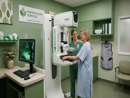
Breast Imaging for Early Detection, Diagnosis & Follow-Up
The Mammography Service at AS Medical Complex provides specialized breast imaging for the screening, investigation, and follow-up of selected breast conditions.
Mammography is a specialized form of medical imaging that uses low-dose X-rays to create detailed images of breast tissue.
A mammogram can help identify abnormalities such as:
-
Breast masses
-
Areas of tissue distortion
-
Calcifications
-
Changes in breast architecture
-
Other suspicious findings
Mammography plays an important role in the early detection of breast cancer because some cancers can be identified on imaging before a patient or healthcare professional can feel a lump.
Mammography is generally divided into two major categories:
-
Screening Mammography
-
Diagnostic Mammography
These examinations have different purposes.
Screening mammography is used in appropriately selected people who do not have breast symptoms.
Diagnostic mammography is used when:
-
A breast symptom is present
-
A clinical abnormality has been identified
-
A screening mammogram requires further evaluation
-
Previous breast imaging requires targeted follow-up
At AS Medical Complex, mammography findings should be interpreted together with:
-
Breast symptoms
-
Clinical examination
-
Age
-
Personal history
-
Family history
-
Previous mammograms
-
Breast density
-
Ultrasound findings where appropriate
-
MRI findings where appropriate
-
Pathology where biopsy has been performed
Mammography is an important diagnostic tool, but an abnormal mammogram alone does not automatically mean breast cancer is present.
Our Mammography Services
Depending on clinical indication, equipment capability, and available breast-imaging expertise, Mammography services at AS Medical Complex may include:
-
Screening Mammography
-
Diagnostic Mammography
-
Bilateral Mammography
-
Unilateral Mammography where clinically appropriate
-
Additional mammographic views
-
Magnification views where indicated
-
Compression views where indicated
-
Evaluation of breast lumps
-
Evaluation of nipple discharge
-
Evaluation of breast skin changes
-
Evaluation of nipple changes
-
Evaluation of clinically detected abnormalities
-
Assessment after an abnormal screening mammogram
-
Follow-up of selected mammographic abnormalities
-
Mammography after previous breast surgery
-
Mammography after breast-conserving treatment where clinically appropriate
-
Mammography in patients with breast implants using appropriate techniques
-
Breast-density assessment
-
Correlation with Breast Ultrasound
-
Correlation with Breast MRI where required
-
Digital Breast Tomosynthesis / 3D Mammography where specifically available
-
Referral for image-guided breast biopsy where required
-
Multidisciplinary coordination with Breast Surgery
-
Follow-up imaging according to radiologist recommendations
Advanced services such as 3D mammography, stereotactic breast biopsy, contrast-enhanced mammography, or other specialized breast procedures should only be advertised as onsite AS Medical Complex services after confirming the relevant equipment, trained personnel, and reporting expertise.
What Is a Mammogram?
A mammogram is an X-ray examination specifically designed to image the breasts.
During the examination:
-
The breast is positioned on a specialized platform.
-
A clear compression paddle gently but firmly compresses the breast.
-
Low-dose X-ray images are obtained.
-
The breast is repositioned for additional views.
Mammography equipment is designed specifically for breast imaging rather than general X-ray examination.
Why Is Mammography Different From a Regular X-Ray?
Breast tissue contains subtle differences between:
-
Fat
-
Fibrous tissue
-
Glandular tissue
-
Masses
-
Calcifications
Mammography equipment and imaging techniques are specifically designed to demonstrate these subtle differences using a low radiation dose.
A routine chest X-ray cannot replace a mammogram for breast-cancer screening or detailed breast assessment.
Screening Mammography
Screening mammography is performed in appropriately selected people who do not have breast symptoms.
Its main purpose is to detect possible breast cancer at an earlier stage, before symptoms appear.
Screening mammography may identify:
-
Small breast masses
-
Suspicious calcifications
-
Architectural distortion
-
Other early imaging changes
RadiologyInfo describes screening mammography as low-dose breast X-ray imaging designed to detect breast cancer before symptoms develop.
Diagnostic Mammography
Diagnostic mammography is performed when a patient has a breast concern or when additional investigation is required.
Possible indications include:
-
Breast lump
-
Nipple discharge
-
Nipple retraction
-
Breast skin dimpling
-
Change in breast shape
-
Focal breast pain in selected circumstances
-
Abnormal screening mammogram
-
Previous imaging requiring further evaluation
Diagnostic mammography may use:
-
Additional views
-
Targeted compression
-
Magnification
-
Other specialized views
to examine a specific area more closely.
Screening Mammogram vs Diagnostic Mammogram
Screening Mammography
Usually performed when:
-
There are no breast symptoms.
-
The purpose is routine early detection.
-
Standard images of both breasts are obtained.
Diagnostic Mammography
Usually performed when:
-
A breast symptom is present.
-
A screening mammogram shows an abnormality.
-
A particular breast area needs closer assessment.
-
Previous breast imaging requires follow-up.
Diagnostic mammography may involve more images and more targeted views than routine screening.
Why Is Early Breast Cancer Detection Important?
Breast cancers discovered at an earlier stage may be smaller and may not yet have spread to lymph nodes or distant organs.
Earlier detection can increase treatment options for appropriately selected patients.
Mammography is therefore an important component of breast-cancer screening programs internationally.
When Should Screening Mammography Begin?
There is no single screening schedule used by every international organization.
For example:
-
The American College of Radiology recommends annual screening beginning at age 40 for women at average risk.
-
The U.S. Preventive Services Task Force recommends screening every two years from ages 40 through 74 for women at average risk.
These recommendations demonstrate that even major international organizations can differ regarding screening intervals.
For patients at AS Medical Complex, screening should therefore consider:
-
Age
-
Personal breast-cancer risk
-
Family history
-
Previous breast disease
-
Genetic risk
-
Previous chest radiation
-
Applicable local clinical recommendations
-
Treating physician advice
Rather than assuming that the same schedule is appropriate for every patient.
Breast Cancer Risk Assessment
A patient’s screening plan may change when breast-cancer risk is higher than average.
Risk factors can include selected:
-
Strong family history
-
Personal history of breast cancer
-
Certain inherited genetic mutations
-
Previous high-risk breast lesions
-
Previous radiation treatment involving the chest at a young age
-
Other significant clinical factors
High-risk patients may require:
-
Earlier screening
-
More frequent mammography
-
Breast MRI
-
Genetic counseling
-
Specialist breast assessment
depending on the individual situation.
Family History of Breast Cancer
A family history may affect breast-cancer risk, especially when cancer occurred in:
-
Mother
-
Sister
-
Daughter
-
Multiple close relatives
or when relatives developed breast or ovarian cancer at younger ages.
However, family history should be evaluated carefully.
Having one relative with breast cancer does not mean that the patient will definitely develop cancer.
A clinician may recommend formal risk assessment when family history is significant.
Genetic Risk
Inherited changes in genes such as BRCA1 and BRCA2 can substantially increase breast-cancer risk in some families.
Patients with:
-
Multiple family members affected
-
Breast cancer at young ages in relatives
-
Ovarian cancer in the family
-
Known familial genetic mutations
may benefit from specialist assessment and genetic counseling.
Mammography alone may not be the complete screening strategy for genetically high-risk patients.
High-Risk Breast Screening
Some high-risk individuals may require both:
-
Mammography
-
Breast MRI
rather than mammography alone.
RadiologyInfo notes that patients at high risk may require breast MRI in addition to mammography.
The screening plan should be individualized through specialist assessment.
Breast Lump
A new breast lump should be medically assessed.
Possible causes include:
-
Cyst
-
Fibroadenoma
-
Infection
-
Fat-related changes
-
Other benign conditions
-
Breast cancer
Depending on age and clinical findings, assessment may involve:
-
Clinical breast examination
-
Ultrasound
-
Diagnostic mammography
-
Biopsy
-
Other imaging
A breast lump should not be ignored simply because it is painless.
Mammography for Breast Lump
Diagnostic mammography may help characterize a breast lump and identify additional abnormalities elsewhere within the breast.
However, mammography may not always determine whether a lump is:
-
Solid
-
Fluid-filled
-
Benign
-
Malignant
Breast Ultrasound may therefore be used alongside mammography.
Breast Ultrasound
Breast Ultrasound uses sound waves rather than X-rays.
It can provide useful information about a breast abnormality identified through:
-
Examination
-
Mammography
-
Another imaging test
Ultrasound can sometimes help distinguish:
-
Simple cyst
-
Solid mass
-
Complex lesion
Mammography and ultrasound frequently provide complementary information rather than one automatically replacing the other.
Mammography vs Breast Ultrasound
Mammography
Particularly useful for:
-
Screening
-
Calcifications
-
Architectural distortion
-
Certain masses
-
Overall breast assessment
Ultrasound
Particularly useful for:
-
Targeted assessment of a lump
-
Distinguishing fluid-filled from solid lesions
-
Evaluating selected abnormalities in younger patients
-
Guiding some biopsies
The radiologist determines whether one or both examinations are required.
Breast MRI
Breast MRI provides high-detail imaging using magnetic fields and radiofrequency energy rather than ionizing radiation.
MRI may be appropriate for selected:
-
High-risk screening
-
Extent-of-disease assessment
-
Implant-related questions
-
Complex breast abnormalities
-
Other specialist indications
Breast MRI is generally not used as a routine replacement for mammography in average-risk patients.
Mammography vs Breast MRI
Mammography may be particularly useful for detecting:
-
Calcifications
-
Small areas of distortion
-
Early screening abnormalities
MRI provides high sensitivity for selected breast lesions but may also detect findings that require additional evaluation.
The best examination depends on the clinical situation.
Nipple Discharge
Nipple discharge may have several possible causes.
Important characteristics include whether discharge is:
-
Spontaneous
-
One-sided
-
Bloody
-
Clear
-
Persistent
-
Coming from one duct
Diagnostic assessment may involve:
-
Clinical examination
-
Mammography
-
Ultrasound
-
MRI in selected circumstances
-
Breast Surgery consultation
A new spontaneous bloody or clear discharge from one breast requires appropriate clinical assessment.
Nipple Retraction
A nipple that has always been inverted may be normal for that individual.
However, new nipple retraction or inversion can require investigation.
Assessment may include:
-
Clinical examination
-
Diagnostic mammography
-
Breast Ultrasound
-
Additional imaging
depending on the patient.
Breast Skin Changes
Breast skin changes requiring medical review may include:
-
Dimpling
-
Thickening
-
Redness
-
Persistent swelling
-
Orange-peel appearance
-
New contour change
Diagnostic mammography may form part of the evaluation, but imaging should not delay clinical assessment when concerning symptoms are present.
Breast Pain
Breast pain is common and most breast pain is not caused by cancer.
Pain may be related to:
-
Hormonal changes
-
Menstrual cycle
-
Musculoskeletal conditions
-
Cysts
-
Infection
-
Other causes
Imaging decisions depend on:
-
Age
-
Focal vs generalized pain
-
Presence of a lump
-
Physical examination
-
Other symptoms
Mammography is not automatically required for every episode of breast tenderness.
Breast Infection
Breast infection or mastitis may cause:
-
Pain
-
Redness
-
Warmth
-
Swelling
-
Fever
Ultrasound is often useful if an abscess is suspected.
Mammography may be appropriate in selected cases, particularly when symptoms persist or there is concern for another underlying breast abnormality.
Breast Abscess
A breast abscess is a collection of infected fluid.
Possible symptoms include:
-
Pain
-
Redness
-
Swelling
-
Fever
-
Localized tender lump
Ultrasound may help confirm the presence of a fluid collection and may assist with drainage planning.
Patients with significant infection require medical treatment rather than imaging alone.
Breast Calcifications
Calcifications are small calcium deposits within breast tissue.
They can appear as white areas on a mammogram.
NCI describes two broad patterns:
Macrocalcifications
Larger calcium deposits that are commonly associated with benign processes such as:
-
Aging
-
Previous injury
-
Inflammation
Microcalcifications
Very small deposits that may occur in benign conditions but can also form patterns associated with ductal carcinoma in situ or breast cancer.
The pattern, shape and distribution are more important than simply whether calcium is present.
Breast Calcium Is Not Caused by Eating Calcium
Mammographic breast calcifications are not the same thing as calcium obtained through food or calcium supplements.
NCI specifically notes that mammographic calcifications are not related to dietary calcium intake.
Patients should not stop medically required calcium solely because calcifications appear on a mammogram.
Breast Mass on Mammography
A mammographic mass may appear with:
-
Smooth borders
-
Irregular borders
-
Round shape
-
Oval shape
-
Other characteristics
Some imaging appearances are more reassuring than others.
However, imaging cannot always establish the diagnosis.
Further assessment may require:
-
Ultrasound
-
Additional mammographic views
-
MRI
-
Biopsy
Architectural Distortion
Architectural distortion refers to an abnormal appearance in the normal arrangement of breast tissue without necessarily forming a clearly defined mass.
It may have benign or malignant causes.
The radiologist may recommend:
-
Additional mammography
-
Tomosynthesis
-
Ultrasound
-
Biopsy
-
Other assessment
depending on its appearance.
Asymmetry
Breast tissue can naturally differ between the two breasts.
An asymmetry on mammography may sometimes represent:
-
Normal overlapping tissue
-
Benign breast tissue
-
A real underlying abnormality
Additional views may be required to determine whether an apparent asymmetry persists.
Additional Mammography Views
A patient may be called back for extra images.
Additional views can include:
-
Spot compression
-
Magnification
-
Different angles
-
Other targeted techniques
The purpose is to obtain a clearer view of an area that was:
-
Partially obscured
-
Uncertain
-
Newly identified
-
Potentially abnormal
Being called back does not automatically mean cancer has been found.
NCI notes that most people called back for additional testing are ultimately not diagnosed with breast cancer.
Magnification Views
Magnification mammography may be used when the radiologist needs a more detailed view of selected abnormalities, particularly:
-
Calcifications
-
Small areas of concern
The technique enlarges the area radiographically so that its features can be assessed more closely.
Spot Compression Views
Spot compression applies focused compression to a specific area of the breast.
It can help determine whether an apparent abnormality represents:
-
Normal overlapping tissue
-
A persistent mass
-
Architectural distortion
-
Another finding
Digital Mammography
Digital mammography uses electronic detectors rather than traditional film.
Images can be:
-
Displayed on computer workstations
-
Magnified
-
Adjusted for viewing
-
Stored digitally
-
Compared with previous studies
RadiologyInfo identifies full-field digital mammography as a modern mammographic technique.
The exact digital mammography equipment installed at AS Medical Complex should be confirmed before equipment-specific claims are published.
3D Mammography / Breast Tomosynthesis
Digital Breast Tomosynthesis – DBT, often called 3D Mammography, obtains multiple low-dose images from different angles and reconstructs them into thin sections through the breast.
This can reduce the problem of overlapping tissue.
RadiologyInfo reports that tomosynthesis can:
-
Improve detection of some breast cancers
-
Reduce some screening callbacks
-
Better demonstrate abnormalities in dense breasts
-
Improve localization of certain findings
3D Mammography should only be listed as an AS Medical Complex service if the installed mammography system specifically supports DBT.
Dense Breasts
Breast density refers to the relative amount of:
-
Fibrous tissue
-
Glandular tissue
-
Fatty tissue
visible on a mammogram.
Dense tissue appears white on mammography.
Many breast cancers also appear white.
This can make some cancers more difficult to identify.
Dense breast tissue is also associated with increased breast-cancer risk.
Breast Density Categories
BI-RADS breast composition is commonly divided into four categories:
-
Almost entirely fatty
-
Scattered areas of fibroglandular density
-
Heterogeneously dense
-
Extremely dense
The final mammography report may describe the patient’s breast density.
Does Dense Breast Tissue Mean Cancer?
No.
Dense breast tissue does not mean that breast cancer is present.
It means:
-
The breast contains more fibroglandular tissue.
-
Mammography may be less sensitive for some cancers.
-
Breast-cancer risk may be somewhat increased.
Whether additional screening is appropriate should be discussed with a healthcare professional based on overall risk.
Supplemental Screening for Dense Breasts
Some patients with dense breasts may benefit from additional imaging depending on their total breast-cancer risk.
Possible additional tests can include:
-
Breast Ultrasound
-
Breast MRI
-
Other specialist breast imaging
Additional imaging should not automatically be ordered for every patient solely because the report says “dense breasts.”
The decision should consider:
-
Age
-
Family history
-
Personal history
-
Genetic risk
-
Degree of density
-
Other risk factors
BI-RADS
Breast imaging reports commonly use the Breast Imaging Reporting and Data System – BI-RADS.
BI-RADS provides standardized terminology and assessment categories for:
-
Mammography
-
Breast Ultrasound
-
Breast MRI
The American College of Radiology updated its BI-RADS manual in 2025 to maintain standardized breast-imaging reporting and management recommendations.
BI-RADS 0
BI-RADS 0 – Incomplete
This generally means the mammogram cannot yet receive a final category because:
-
Additional imaging is needed
-
Previous imaging may be required for comparison
BI-RADS 0 does not mean cancer has been diagnosed.
BI-RADS 1
BI-RADS 1 – Negative
No suspicious abnormality is identified.
Routine screening is generally recommended according to the patient’s appropriate schedule.
BI-RADS 2
BI-RADS 2 – Benign
A benign finding is present.
Examples may include selected:
-
Cysts
-
Calcifications
-
Other clearly non-cancerous findings
Routine screening is generally recommended.
BI-RADS 3
BI-RADS 3 – Probably Benign
The finding has a very low likelihood of cancer but usually requires short-interval imaging follow-up.
A six-month follow-up examination is commonly recommended.
BI-RADS 4
BI-RADS 4 – Suspicious
A suspicious abnormality is present.
Biopsy is generally recommended.
BI-RADS 4 can be subdivided into:
-
4A – lower suspicion
-
4B – moderate suspicion
-
4C – higher suspicion
A BI-RADS 4 result still does not mean cancer has definitely been diagnosed.
The diagnosis requires tissue sampling.
BI-RADS 5
BI-RADS 5 – Highly Suggestive of Malignancy
The imaging appearance is highly suspicious for breast cancer.
Biopsy is strongly recommended to establish the diagnosis.
BI-RADS 6
BI-RADS 6 – Known Biopsy-Proven Malignancy
This category is used when cancer has already been confirmed by biopsy and imaging is being performed in that known clinical context.
BI-RADS Is Not the Same as Cancer Stage
BI-RADS communicates the level of imaging concern and recommended next step.
It does not represent breast-cancer stage.
Cancer stage is determined using information such as:
-
Tumor size
-
Lymph-node involvement
-
Spread to other organs
-
Pathology
-
Other clinical investigations
Abnormal Mammogram Does Not Automatically Mean Cancer
An abnormal mammogram can result from many conditions.
Possible benign explanations include:
-
Cysts
-
Fibroadenomas
-
Benign calcifications
-
Previous surgery
-
Scar tissue
-
Normal tissue overlap
-
Other non-cancerous changes
Further imaging or biopsy is used to establish the diagnosis when necessary.
Mammography Cannot Confirm Cancer Alone
Mammography can identify suspicious abnormalities, but the definitive diagnosis of breast cancer generally requires biopsy and pathology.
NCI notes that mammography can identify abnormal tissue but biopsy is required to determine whether suspicious tissue is cancerous.
Breast Biopsy
A breast biopsy removes tissue or cells from a suspicious area for laboratory examination.
Depending on the lesion, biopsy may be performed using:
-
Ultrasound guidance
-
Mammographic/stereotactic guidance
-
MRI guidance
-
Surgical techniques
The appropriate method depends on:
-
Lesion location
-
Imaging visibility
-
Size
-
Other clinical considerations
Mammography & Breast Surgery
The Breast Surgery Department may become involved when mammography or other imaging identifies:
-
Suspicious breast lesion
-
Biopsy-proven cancer
-
High-risk breast abnormality
-
Persistent breast symptoms
-
Other surgical breast conditions
Breast imaging, Pathology and Surgery should work together to establish the correct diagnosis and treatment plan.
Mammography After Breast Surgery
Previous breast surgery can change the appearance of breast tissue.
Patients should inform the mammography team about previous:
-
Breast biopsy
-
Lumpectomy
-
Breast-conserving surgery
-
Reduction surgery
-
Reconstruction
-
Implant surgery
-
Other breast procedures
RadiologyInfo recommends informing the imaging team about prior breast surgery because it can affect mammographic interpretation.
Mammography After Breast Cancer Treatment
Patients who have undergone breast-conserving treatment may require ongoing mammographic surveillance according to their specialist follow-up plan.
The schedule depends on:
-
Type of treatment
-
Surgery
-
Radiation therapy
-
Remaining breast tissue
-
Previous imaging findings
-
Oncology/Breast Surgery recommendations
Follow-up should be individualized.
Previous Mastectomy
Routine mammography is generally performed on breast tissue that remains.
Patients who have undergone mastectomy require individualized surveillance according to:
-
Type of surgery
-
Reconstruction
-
Symptoms
-
Specialist recommendations
Any new lump or change involving the chest wall or reconstruction should be clinically assessed.
Breast Implants
Patients with breast implants can still require mammography.
However, implants can obscure some breast tissue on standard mammographic views.
RadiologyInfo notes that both saline and silicone implants can block visualization of surrounding breast tissue.
Patients should always tell the mammography facility that they have implants when scheduling the examination.
Implant-Displacement Views
Additional specialized views may be used in patients with breast implants to improve visualization of breast tissue.
The technologist may carefully reposition the breast tissue relative to the implant.
The exact technique depends on:
-
Implant location
-
Breast anatomy
-
Surgery
-
Patient tolerance
Can Compression Rupture a Breast Implant?
Mammography technologists use modified techniques when implants are present.
Implant rupture during mammography is uncommon, but patients should:
-
Tell the technologist about their implants.
-
Report any known implant problem.
-
Provide relevant surgical history.
RadiologyInfo notes that experienced technologists know how to compress and position the breast carefully in patients with implants.
Mammography Does Not Fully Evaluate Implant Integrity
Mammography is primarily designed to evaluate breast tissue.
When there is concern about the integrity of a breast implant, particularly a silicone implant, additional imaging such as:
-
Ultrasound
-
MRI
may be recommended depending on the situation.
Mammography in Men
Men can also develop breast cancer, although it is much less common than in women.
A man with symptoms such as:
-
New breast lump
-
Nipple retraction
-
Bloody nipple discharge
-
Significant unilateral breast change
should undergo medical assessment.
Diagnostic mammography and/or ultrasound may be used in selected male patients.
Routine population mammography screening is not normally performed in average-risk men.
Gynecomastia
Gynecomastia is benign enlargement of male breast tissue.
Possible causes can include:
-
Hormonal changes
-
Medication
-
Liver disease
-
Other medical conditions
Imaging may be appropriate when clinical findings are uncertain or suspicious.
Preparing for a Mammogram
Mammography generally requires little preparation.
Patients should:
-
Bring previous mammograms where possible.
-
Tell staff about breast symptoms.
-
Tell staff about previous breast surgery.
-
Tell staff about implants.
-
Tell staff if pregnancy is possible.
-
Follow the imaging facility’s specific instructions.
Previous mammograms are particularly valuable because comparison can help the radiologist identify whether a finding is:
-
New
-
Stable
-
Changing
Do Not Use Deodorant or Powder
On the day of a mammogram, patients may be asked to avoid products such as:
-
Deodorant
-
Antiperspirant
-
Talcum powder
-
Lotion
-
Cream
on the breasts or underarms.
Particles from some products can appear on the mammogram and potentially interfere with image interpretation.
Clothing for Mammography
It may be more convenient to wear a two-piece outfit because the patient usually needs to remove clothing from the upper body.
A gown may be provided.
Jewelry near the imaging area may also need to be removed.
Menstrual Cycle & Breast Tenderness
Some patients experience greater breast tenderness shortly before or during menstruation.
If mammography is being performed routinely and there is flexibility in scheduling, choosing a time when the breasts are less tender may improve comfort.
RadiologyInfo suggests avoiding the week before menstruation for patients whose breasts are usually especially tender during that period.
A medically necessary diagnostic examination should not be unnecessarily delayed because of the menstrual cycle.
Pregnancy & Mammography
Patients should always tell the doctor and mammography technologist if:
-
They are pregnant.
-
They think pregnancy is possible.
Routine screening mammography is generally approached differently during pregnancy.
However, a concerning breast symptom during pregnancy should still be investigated.
The imaging team may use:
-
Ultrasound
-
Mammography when clinically necessary
-
Other appropriate assessment
depending on the situation.
Because the X-ray beam is directed at the breast and mammography uses a low dose, diagnostic mammography may still be considered when the expected medical benefit justifies the examination.
The decision should be individualized.
Breast Symptoms During Pregnancy
Pregnancy can cause many normal breast changes.
However, a persistent:
-
Lump
-
Bloody discharge
-
Focal skin change
-
Other concerning abnormality
should not simply be attributed to pregnancy without clinical assessment.
Ultrasound is often an important first imaging test during pregnancy.
Additional imaging may be required depending on findings.
Mammography During Breastfeeding
Breastfeeding does not automatically prevent medically necessary breast imaging.
However, lactation can increase breast density.
For comfort and potentially improved imaging, a breastfeeding patient may be advised to:
-
Feed the baby
-
Pump milk
shortly before the examination.
The radiology team should be informed that the patient is breastfeeding.
What Happens During a Mammogram?
During mammography:
-
Patient identity is confirmed.
-
Relevant history is reviewed.
-
The patient removes clothing from the upper body.
-
One breast is positioned on the mammography platform.
-
A compression paddle is lowered.
-
The breast is compressed for a few seconds.
-
The image is obtained.
-
The breast is repositioned for another view.
-
The process is repeated for the other breast where required.
Screening mammography typically uses standard views, while diagnostic mammography may require additional images.
Why Is Breast Compression Necessary?
Compression is an important part of mammography.
It helps:
-
Spread breast tissue
-
Reduce tissue overlap
-
Improve visibility of small abnormalities
-
Reduce movement
-
Improve image sharpness
-
Reduce the amount of radiation required
RadiologyInfo specifically identifies these reasons for breast compression during mammography.
Is Mammography Painful?
Most patients experience pressure during compression.
Some find it:
-
Mildly uncomfortable
-
Moderately uncomfortable
-
Occasionally painful
The compression generally lasts only a short time for each image.
FDA notes that while some patients find the pressure uncomfortable, it usually lasts only a few seconds.
Tell the technologist if the discomfort becomes severe.
Why Can't the Breast Be Imaged Without Compression?
Without adequate compression:
-
Breast tissue overlaps more.
-
Small abnormalities may be hidden.
-
Motion blur can increase.
-
More radiation may be required.
-
Image quality can decrease.
Compression therefore improves both image quality and radiation efficiency.
How Long Does a Mammogram Take?
The exact appointment duration depends on:
-
Screening vs diagnostic examination
-
Number of views
-
Need for additional imaging
-
Breast implants
-
Mobility
-
Previous surgery
-
Other factors
The actual X-ray exposure for each image is very brief.
Diagnostic mammography may take longer because more views may be required.
AS Medical Complex should avoid promising one fixed examination time for every patient.
Radiation Dose From Mammography
Mammography uses low-dose ionizing radiation.
Like all X-ray-based imaging, radiation carries a small potential risk.
However, FDA states that the radiation risk associated with mammography is very small compared with the benefit of detecting breast cancer early in appropriate screening populations.
The examination should still be:
-
Clinically justified
-
Performed using appropriate breast-imaging equipment
-
Conducted according to quality-control procedures
Does Radiation Stay in the Body?
No.
The X-rays used during mammography pass through the body to create the image.
No X-ray radiation remains inside the patient afterward.
The patient does not become radioactive.
Mammography Quality
Reliable mammography depends on more than simply having an X-ray machine.
Quality involves:
-
Dedicated breast-imaging equipment
-
Proper positioning
-
Adequate compression
-
Appropriate radiation exposure
-
Trained technologists
-
Appropriate radiologist interpretation
-
Equipment quality control
-
Comparison with prior imaging
-
Clear follow-up recommendations
AS Medical Complex should advertise any specific accreditation or certification only after it has been formally obtained and verified.
Importance of Previous Mammograms
Comparison with previous mammograms can be extremely valuable.
A finding that appears concerning on one image may have been:
-
Present for many years
-
Completely stable
-
Previously investigated
Conversely, comparison may reveal a subtle new change.
RadiologyInfo recommends bringing previous breast imaging to allow comparison whenever possible.
Mammography Results
A mammography report may include:
-
Reason for examination
-
Imaging technique
-
Breast density
-
Findings
-
Comparison with previous mammograms
-
BI-RADS assessment
-
Recommendation
Patients should review the report with the healthcare professional responsible for their breast care.
Normal Mammogram Does Not Exclude Every Breast Cancer
Mammography is highly valuable but is not perfect.
Some cancers may be difficult to see because of:
-
Dense breast tissue
-
Tumor location
-
Breast anatomy
-
Previous surgery
-
Implants
-
Other factors
RadiologyInfo states clearly that not all breast cancers can be visualized on mammography.
A patient with a persistent concerning breast lump or other symptom requires clinical evaluation even if a recent mammogram was reported as normal.
False-Negative Mammogram
A false-negative mammogram occurs when breast cancer is present but is not identified on the mammogram.
This is one reason why mammography should not replace:
-
Clinical assessment
-
Appropriate ultrasound
-
Biopsy
-
Other imaging
when a significant physical abnormality persists.
False-Positive Mammogram
A false-positive result occurs when mammography identifies an abnormality that ultimately proves not to be cancer.
This may lead to:
-
Additional mammographic views
-
Ultrasound
-
Biopsy
-
Short-term follow-up
Callbacks and additional testing are an expected part of breast screening, and most people called back do not ultimately have breast cancer.
Callback After Mammography
Being called back for additional imaging means that more information is needed.
Possible reasons include:
-
Tissue overlap
-
Calcifications
-
Asymmetry
-
Mass
-
Distortion
-
Need to compare prior studies
Patients should attend recommended follow-up promptly rather than assuming that a callback either definitely means cancer or can safely be ignored.
Short-Interval Follow-Up
Certain findings categorized as probably benign may require repeat imaging after a shorter interval rather than immediate biopsy.
BI-RADS 3 findings commonly receive approximately six-month imaging follow-up.
The purpose is to confirm stability.
Mammography & Breast Cancer Diagnosis
When imaging is suspicious, the diagnostic pathway may include:
-
Diagnostic mammography
-
Breast Ultrasound
-
Breast MRI
-
Biopsy
-
Pathology
-
Breast Surgery consultation
The final diagnosis of cancer is established by tissue evaluation rather than mammography alone.
Mammography & Breast Surgery Department
The Breast Surgery team may evaluate patients with:
-
Suspicious mammography
-
Biopsy-proven breast cancer
-
Persistent breast lump
-
Nipple discharge
-
Recurrent breast symptoms
-
High-risk lesions
-
Other surgical breast conditions
Close coordination between:
-
Radiology
-
Breast Surgery
-
Pathology
-
Oncology
can support appropriate diagnosis and treatment planning.
Mammography & Oncology
Once breast cancer has been confirmed, Oncology management may require information from:
-
Mammography
-
Ultrasound
-
MRI
-
Biopsy
-
Pathology
-
Surgical findings
-
Other staging investigations
The imaging strategy depends on cancer type and stage.
Mammography & Gynecology / Well Women Clinic
The Well Women Clinic and Gynecology services may help identify patients who require breast-health assessment based on:
-
Age
-
Symptoms
-
Family history
-
Menopausal status
-
Other risk factors
Screening decisions should be individualized rather than based on symptoms alone.
Mammography & Radiology
Mammography forms part of the broader Radiology Service at AS Medical Complex, but breast imaging requires specialized techniques and interpretation.
Radiology may coordinate:
-
Mammography
-
Breast Ultrasound
-
Breast MRI
-
Additional diagnostic imaging
-
Image-guided biopsy referral where necessary
depending on findings.
Thermography Is Not a Replacement for Mammography
Thermography measures heat patterns but has not been established as an adequate replacement for mammography for breast-cancer screening.
FDA specifically states that thermography should not be used as a substitute for mammography.
Patients should be cautious about services claiming that thermography can replace evidence-based breast imaging.
Mammography Is Not a Blood Test for Cancer
There is currently no routine blood test that replaces mammography for standard breast-cancer screening.
Likewise, tumor-marker tests should not be used as substitutes for appropriate breast imaging in otherwise asymptomatic screening.
Breast-cancer detection requires an appropriate combination of:
-
Risk assessment
-
Imaging
-
Clinical evaluation
-
Biopsy when necessary
Mammography Limitations
Mammography has several limitations.
These include:
-
Some cancers may not be visible.
-
Dense tissue can reduce sensitivity.
-
False-positive findings can occur.
-
Breast implants may obscure tissue.
-
Some abnormalities require ultrasound or MRI.
-
Mammography cannot provide a tissue diagnosis.
-
Additional imaging may be required.
These limitations do not eliminate the value of mammography but explain why breast care often involves more than one diagnostic method.
When Should You Consider a Screening Mammogram?
Screening mammography may be appropriate when:
-
You have reached an age at which screening is recommended according to your risk and applicable clinical guidance.
-
You have no breast symptoms but require routine screening.
-
Your doctor recommends mammography based on your risk profile.
-
You have a higher-than-average breast-cancer risk requiring an individualized surveillance plan.
Screening frequency should be discussed with your healthcare professional.
When Should You Have Diagnostic Breast Assessment Instead?
Do not simply book a routine screening mammogram when you have a new breast symptom.
Inform the healthcare team if you have:
-
New breast lump
-
New nipple discharge
-
Nipple retraction
-
Breast skin dimpling
-
New focal breast change
-
Persistent one-sided breast abnormality
-
Previous abnormal breast imaging
You may require diagnostic mammography and/or ultrasound rather than a routine screening examination.
When Should a Breast Lump Be Assessed?
Seek medical assessment if you notice:
-
New breast lump
-
Thickened area
-
Lump in the armpit
-
Persistent change from your normal breast tissue
Do not wait for the next routine screening date if you have a new concerning symptom.
When Is Urgent Medical Assessment Required?
Most breast conditions are evaluated through outpatient breast clinics rather than Emergency care.
However, urgent medical review may be required when there is:
-
Severe rapidly spreading breast infection
-
High fever with breast redness and systemic illness
-
Rapidly increasing painful swelling
-
Significant postoperative bleeding
-
Severe complication after breast surgery
-
Other acute medical deterioration
A suspected breast cancer usually requires prompt specialist outpatient evaluation, but is not typically an Emergency Department condition unless an acute complication is present.
Multidisciplinary Breast Care
Depending on mammography findings, patient care may involve:
-
Radiology
-
Breast Surgery
-
General Surgery
-
Oncology
-
Pathology
-
Gynecology
-
Well Women Clinic
-
Plastic Surgery
-
Other appropriate specialties
The goal is to move efficiently from:
-
Screening
-
Detection
-
Diagnostic imaging
-
Biopsy
-
Diagnosis
-
Treatment planning
when necessary.
Follow-Up After Mammography
The next step depends on the mammography findings.
Possible recommendations include:
-
Routine screening
-
Previous-image comparison
-
Additional mammography views
-
Breast Ultrasound
-
Short-interval follow-up
-
Breast MRI
-
Biopsy
-
Breast Surgery consultation
-
Other specialist assessment
Patients should follow the recommendation written in the radiology report.
Do Not Ignore Mammography Results
Patients should ensure that they receive their mammography report.
RadiologyInfo specifically advises patients not to assume that results are normal simply because they have not been contacted.
If a recommended follow-up examination is advised, it should be arranged within the appropriate timeframe.
Long-Term Breast Imaging Follow-Up
Breast health is not based on a single mammogram.
Depending on risk and previous findings, long-term care may include:
-
Regular screening
-
Comparison with prior studies
-
Short-term surveillance of selected findings
-
Ultrasound
-
MRI in higher-risk patients
-
Clinical examination
-
Specialist follow-up
Using previous imaging consistently can make subtle changes easier to identify.
Mammography FAQs
What is a mammogram?
A mammogram is a specialized low-dose X-ray examination of the breasts. Mammography is used both for breast-cancer screening in appropriately selected people without symptoms and for diagnostic assessment of symptoms or abnormal screening findings. It can identify masses, calcifications and other breast changes that may not yet be noticeable on physical examination.
What is the difference between screening and diagnostic mammography?
Screening mammography is performed for early breast-cancer detection in people without breast symptoms. Diagnostic mammography is used when there is a breast symptom—such as a lump or nipple discharge—or when a screening mammogram identifies an area that requires further evaluation. Diagnostic mammography may require more targeted images and additional views.
Is mammography painful?
The breast must be compressed to spread the tissue, reduce overlap, minimize movement and obtain a clear image using a lower radiation dose. Most patients experience pressure or temporary discomfort, while some find it painful. The compression for each image usually lasts only a few seconds.
Does mammography use radiation?
Yes. Mammography uses a low dose of ionizing X-ray radiation. The radiation risk is considered very small compared with the potential benefit of detecting breast cancer earlier when mammography is appropriately used. No radiation remains in the body after the examination.
What does BI-RADS mean on a mammogram report?
BI-RADS is the Breast Imaging Reporting and Data System used to standardize mammography, breast-ultrasound and breast-MRI reporting. Categories range from BI-RADS 0, meaning additional information or imaging is needed, through categories describing negative, benign, probably benign or suspicious findings, to BI-RADS 6 for known biopsy-proven cancer. A BI-RADS category is an imaging assessment—not automatically a cancer diagnosis.
What should I do before a mammogram?
Bring previous mammograms whenever possible and tell the imaging team about breast symptoms, previous breast surgery, implants, family or personal breast-cancer history, and possible pregnancy. On the day of the examination, avoid deodorant, antiperspirant, powder, lotion or cream on the breasts and underarms because these products may interfere with image interpretation.
Does an abnormal mammogram mean that I have breast cancer?
No. Many abnormal mammograms are caused by benign conditions, normal tissue overlap, cysts, calcifications or other non-cancerous changes. Additional mammography, ultrasound, MRI or biopsy may be recommended depending on the finding. Mammography can identify suspicious tissue, but biopsy and Pathology are generally required to confirm whether cancer is present.
Mammography Services at AS Medical Complex
The Mammography Service at AS Medical Complex is designed to support the early detection, investigation and follow-up of breast disease through clinically appropriate specialized breast imaging.
From screening mammography and diagnostic mammography to assessment of breast lumps, nipple discharge, breast changes, suspicious calcifications, breast density, abnormal screening findings and selected postoperative breast follow-up, our goal is to provide imaging information that supports timely and informed breast-care decisions.
Mammography uses low-dose X-rays and remains an important primary imaging method for breast-cancer screening. However, mammography should be understood as one part of a complete breast diagnostic pathway.
Depending on the patient’s symptoms and mammography findings, additional assessment may include:
-
Breast Ultrasound
-
Breast MRI
-
Additional diagnostic mammographic views
-
Short-term imaging follow-up
-
Breast biopsy
-
Pathology
-
Breast Surgery consultation
-
Oncology consultation
Patients with breast implants, previous breast surgery, dense breast tissue, significant family history, or other high-risk factors may require an individualized imaging approach.
AS Medical Complex should confirm the availability of Digital Breast Tomosynthesis / 3D Mammography, image-guided biopsy and other specialized breast-imaging procedures before these are promoted as onsite services.
Mammography findings can be coordinated with relevant services at AS Medical Complex, including Radiology, Breast Surgery, General Surgery, Gynecology, Well Women Clinic, Pathology, Oncology, Plastic Surgery and other appropriate specialties.
Book a Mammography appointment at AS Medical Complex for physician-recommended breast-cancer screening, diagnostic assessment of breast symptoms, follow-up of abnormal breast imaging, and other clinically indicated breast-imaging examinations.