CT Scan at AS Medical Complex
Contact hospital
- Availability
Contact hospital
- Booking
WhatsApp / call
- Location
Islamabad
Computed Tomography for Rapid & Detailed Diagnostic Imaging
The CT Scan Service at AS Medical Complex provides detailed diagnostic imaging for patients who require rapid assessment of internal organs, bones, blood vessels, soft tissues, injuries, infections, tumors, and other medical conditions.
Computed Tomography – commonly known as a CT Scan or CAT Scan – uses specialized X-ray equipment and computer processing to create detailed cross-sectional images of the body.
These images can show internal structures in much greater detail than many conventional X-rays.
CT can be particularly useful for examining:
-
Brain
-
Head
-
Facial bones
-
Sinuses
-
Chest
-
Lungs
-
Abdomen
-
Pelvis
-
Kidneys
-
Urinary tract
-
Bones
-
Spine
-
Blood vessels
-
Soft tissues
-
Other internal structures
CT is particularly valuable in emergency medicine because images can often be obtained quickly.
For example, CT may help assess:
-
Head injury
-
Internal bleeding
-
Stroke-related conditions
-
Major trauma
-
Severe abdominal pain
-
Kidney stones
-
Lung disease
-
Selected vascular emergencies
MedlinePlus describes CT as an imaging technique using specialized X-ray equipment to create detailed cross-sectional pictures of internal organs, bones, and other tissues.
At AS Medical Complex, CT examinations should be selected according to the patient’s symptoms, medical history, suspected diagnosis, previous investigations, radiation considerations, contrast requirements, and specialist or radiologist recommendation.
Our CT Scan Services
Depending on clinical indication, available CT protocols, and specialist recommendation, CT services at AS Medical Complex may include:
-
CT Brain
-
CT Head
-
CT Facial Bones
-
CT Sinuses
-
CT Neck
-
CT Chest
-
CT Lungs
-
CT Abdomen
-
CT Pelvis
-
CT Abdomen & Pelvis
-
CT Kidneys, Ureters & Bladder where indicated
-
CT for kidney stones
-
CT Spine
-
CT Cervical Spine
-
CT Thoracic Spine
-
CT Lumbar Spine
-
CT Bones
-
CT for trauma
-
CT for selected fractures
-
CT for selected infections
-
CT for selected tumors
-
CT for cancer assessment
-
CT for treatment follow-up
-
CT for postoperative assessment
-
CT without contrast
-
Contrast-enhanced CT where clinically indicated
-
Selected CT Angiography where available
-
Selected pulmonary CT angiography where available
-
Selected vascular CT studies
-
Selected high-resolution chest CT protocols where available
-
Selected low-dose CT protocols where clinically appropriate
-
Pediatric CT where medically indicated
-
Emergency CT imaging
-
Inpatient CT imaging
-
Specialist CT review
-
Follow-up CT
-
Referral for advanced CT protocols where required
Advanced CT protocols should only be advertised as onsite services after confirming the scanner capability, contrast-injection facilities, software, monitoring arrangements, and reporting expertise available at AS Medical Complex.
What Is a CT Scan?
A CT scan uses X-rays taken from multiple angles around the patient.
A computer processes these measurements to create images representing thin sections or “slices” through the body.
These images allow doctors to examine internal structures in detail.
CT images can also be reconstructed into:
-
Different anatomical planes
-
Three-dimensional images
-
Vascular images
-
Bone reconstructions
depending on the examination.
FDA describes CT as a technique in which an X-ray source rotates around the patient while computer processing creates cross-sectional images of internal anatomy.
CT Uses X-Ray Radiation
Unlike MRI and ultrasound, CT uses ionizing radiation.
The amount of radiation depends on factors such as:
-
Body area being scanned
-
Patient size
-
Scan protocol
-
Number of scan phases
-
Clinical indication
-
Equipment settings
FDA notes that CT examinations involve ionizing radiation and that this exposure may produce a small increase in lifetime cancer risk. However, when CT is medically justified, the diagnostic benefit generally outweighs this small potential risk.
This is why CT should be performed when it is expected to provide clinically useful information.
CT vs Conventional X-Ray
A conventional X-ray generally produces a two-dimensional projection image.
CT produces multiple cross-sectional images.
For example, an X-ray may be useful for:
-
Many fractures
-
Chest assessment
-
Joint alignment
-
Certain bone conditions
CT can provide much greater anatomical detail for:
-
Complex fractures
-
Internal organs
-
Brain
-
Internal bleeding
-
Lungs
-
Abdominal disease
-
Blood vessels
-
Other structures
CT is not automatically required whenever an X-ray is abnormal.
The appropriate next investigation depends on the clinical question.
CT vs MRI
CT and MRI are both important imaging methods but have different strengths.
CT Scan
CT is often particularly useful for:
-
Emergency assessment
-
Major trauma
-
Acute head injury
-
Suspected intracranial bleeding
-
Lung imaging
-
Complex fractures
-
Kidney stones
-
Acute abdominal disease
-
Selected vascular conditions
CT examinations are generally relatively fast.
MRI
MRI can provide particularly detailed evaluation of many:
-
Brain structures
-
Spinal cord abnormalities
-
Nerves
-
Ligaments
-
Tendons
-
Muscles
-
Soft tissues
-
Joint structures
MRI does not use ionizing radiation.
FDA notes that MRI generally provides greater soft-tissue contrast than CT, whereas CT is often particularly effective for bone and rapid imaging.
The correct investigation depends on the clinical situation rather than one modality always being superior.
CT vs Ultrasound
Ultrasound and CT also answer different medical questions.
Ultrasound may be particularly useful for:
-
Gallbladder
-
Pregnancy
-
Pelvic assessment
-
Thyroid
-
Superficial structures
-
Blood-flow studies
-
Selected abdominal conditions
CT may provide a broader and more detailed overview of:
-
Abdomen
-
Pelvis
-
Chest
-
Trauma
-
Deep internal structures
-
Complex disease
Ultrasound does not use ionizing radiation, while CT does.
Doctors therefore consider both diagnostic value and radiation exposure when selecting imaging.
CT Brain
CT of the brain is one of the most frequently used emergency imaging examinations.
A head CT may be used to evaluate selected patients with:
-
Head injury
-
Suspected intracranial bleeding
-
Sudden neurological symptoms
-
Selected stroke presentations
-
Severe headache with concerning features
-
Loss of consciousness
-
Certain seizures
-
Suspected brain mass
-
Hydrocephalus
-
Skull abnormalities
-
Other neurological conditions
RadiologyInfo notes that head CT can rapidly identify injuries and intracranial bleeding and can help evaluate conditions involving stroke, aneurysm, tumors, and other neurological abnormalities.
CT After Head Injury
CT can be particularly valuable following significant head trauma.
It may help identify:
-
Skull fracture
-
Intracranial bleeding
-
Brain swelling
-
Other traumatic injury
Not every minor head injury requires CT.
The decision depends on factors such as:
-
Loss of consciousness
-
Neurological symptoms
-
Vomiting
-
Age
-
Use of blood thinners
-
Mechanism of injury
-
Clinical examination
-
Other risk factors
Unnecessary CT should be avoided when imaging is unlikely to change treatment.
CT & Stroke
A patient with possible stroke requires urgent Emergency assessment.
Symptoms may include:
-
Sudden facial weakness
-
Sudden arm weakness
-
Sudden leg weakness
-
Sudden speech difficulty
-
Sudden loss of coordination
-
Sudden visual disturbance
Emergency brain imaging may include:
-
Non-contrast CT
-
CT angiography
-
CT perfusion in selected settings
-
MRI
-
Other vascular imaging
depending on the clinical pathway.
CT can be particularly valuable because it can rapidly assess for intracranial bleeding.
A patient with suspected stroke should not wait for a routine outpatient CT appointment.
Intracranial Bleeding
Bleeding inside the skull can become life-threatening.
CT may help identify:
-
Intracerebral hemorrhage
-
Subdural bleeding
-
Epidural bleeding
-
Subarachnoid hemorrhage
-
Other acute intracranial blood
Further treatment may require:
-
Neurology
-
Neurosurgery
-
Critical Care
-
Other emergency services
depending on the findings.
Headache & CT
Most headaches do not automatically require CT.
The doctor may consider imaging when headache is associated with concerning features such as:
-
Sudden severe onset
-
Neurological abnormalities
-
Head injury
-
Loss of consciousness
-
Fever with concerning neurological signs
-
Significant change in usual headache pattern
-
Other clinical warning signs
The appropriate investigation may be CT or MRI depending on the circumstances.
CT Facial Bones
CT can provide detailed assessment of the bones of the face.
Possible indications include:
-
Facial trauma
-
Suspected facial fracture
-
Complex injury
-
Selected tumors
-
Other abnormalities
Three-dimensional reconstructions may sometimes help surgical planning.
CT Sinuses
CT can provide detailed imaging of:
-
Paranasal sinuses
-
Nasal passages
-
Surrounding bony anatomy
CT of the sinuses may be considered in selected patients with:
-
Chronic sinusitis
-
Complicated sinus disease
-
Nasal polyps
-
Surgical planning
-
Other ENT indications
Routine CT is not required for every episode of uncomplicated sinus symptoms.
The ENT specialist should determine when imaging is appropriate.
CT Neck
CT of the neck may help evaluate selected conditions involving:
-
Deep neck tissues
-
Lymph nodes
-
Airway
-
Salivary glands
-
Thyroid surroundings
-
Infection
-
Masses
-
Trauma
Contrast may be useful in many neck CT examinations.
The appropriate protocol depends on the suspected disease.
CT Chest
CT of the chest provides detailed images of structures including:
-
Lungs
-
Airways
-
Pleura
-
Mediastinum
-
Chest wall
-
Selected blood vessels
-
Other thoracic structures
It may be used to assess:
-
Lung nodules
-
Lung masses
-
Pneumonia complications
-
Interstitial lung disease
-
Pleural disease
-
Selected infections
-
Trauma
-
Other chest abnormalities
CT can show much more anatomical detail than a routine chest X-ray.
Lung Nodules
A lung nodule is a small abnormal area within the lung.
Many lung nodules are benign.
Possible causes include:
-
Previous infection
-
Inflammation
-
Scar tissue
-
Benign growths
-
Cancer
Management depends on:
-
Size
-
Appearance
-
Growth over time
-
Patient age
-
Smoking history
-
Cancer history
-
Other risk factors
Some nodules require follow-up CT, while others may not require additional action.
CT & Lung Cancer
CT plays an important role in:
-
Evaluation of suspicious lung abnormalities
-
Tumor assessment
-
Staging
-
Treatment planning
-
Follow-up
However, a suspicious CT finding does not automatically confirm cancer.
Diagnosis may require:
-
Specialist consultation
-
PET imaging where appropriate
-
Bronchoscopy
-
CT-guided biopsy where available
-
Surgery
-
Histopathology
-
Other investigations
Low-Dose CT Lung Screening
Low-dose CT is used in some evidence-based lung-cancer screening programs for carefully selected people at high risk, particularly based on age and significant smoking exposure.
This differs from performing a routine chest CT on everyone.
Eligibility should be determined according to applicable screening guidelines and individual risk.
Availability of a formal low-dose lung-cancer screening program at AS Medical Complex should be confirmed before it is advertised.
High-Resolution CT – HRCT
High-resolution CT protocols may provide detailed assessment of lung tissue.
HRCT can be useful in selected patients with suspected:
-
Interstitial lung disease
-
Pulmonary fibrosis
-
Bronchiectasis
-
Other diffuse lung abnormalities
Pulmonology and Radiology teams may coordinate the appropriate protocol.
Availability should be confirmed according to scanner capability.
Pneumonia & CT
Most uncomplicated pneumonia does not automatically require CT.
Chest X-ray and clinical examination may be sufficient initially.
CT may be considered when:
-
Diagnosis remains uncertain
-
Complication is suspected
-
Patient is not improving as expected
-
Abscess or empyema is suspected
-
Another underlying condition is being considered
The treating doctor determines the appropriate imaging pathway.
CT Pulmonary Angiography
CT Pulmonary Angiography – CTPA is a contrast-enhanced CT examination designed to evaluate pulmonary blood vessels.
One important indication is suspected pulmonary embolism – PE, where a blood clot blocks blood flow within the lungs.
Patients with symptoms such as:
-
Sudden severe breathlessness
-
Chest pain
-
Low oxygen level
-
Collapse
-
Other signs of possible PE
require urgent clinical assessment.
CTPA should be performed according to an appropriate emergency or specialist pathway.
Availability at AS Medical Complex should be confirmed before advertising it as a routine onsite service.
CT Abdomen
CT of the abdomen can provide detailed images of structures such as:
-
Liver
-
Gallbladder region
-
Pancreas
-
Spleen
-
Kidneys
-
Adrenal glands
-
Bowel
-
Blood vessels
-
Lymph nodes
-
Other abdominal tissues
Body CT can help detect conditions involving infection, inflammation, trauma, tumors, internal bleeding, and many other abdominal abnormalities.
Abdominal Pain & CT
Abdominal pain has many possible causes.
CT may be appropriate in selected patients with suspected:
-
Appendicitis
-
Bowel obstruction
-
Diverticulitis
-
Abscess
-
Perforation
-
Kidney stones
-
Internal bleeding
-
Trauma
-
Tumor
-
Other acute abdominal conditions
However, CT is not required for every episode of abdominal discomfort.
Depending on the patient, the first investigation may instead be:
-
Ultrasound
-
Laboratory testing
-
Endoscopy
-
Clinical observation
-
Another examination
Appendicitis
Appendicitis can cause:
-
Abdominal pain
-
Pain moving toward the right lower abdomen
-
Fever
-
Nausea
-
Loss of appetite
-
Other symptoms
CT can help diagnose appendicitis in selected patients when imaging is required.
Ultrasound may sometimes be preferred initially, particularly in:
-
Children
-
Younger patients
-
Pregnancy
depending on clinical circumstances and available expertise.
Bowel Obstruction
CT can help assess suspected bowel obstruction.
It may provide information about:
-
Location of obstruction
-
Degree of obstruction
-
Bowel dilation
-
Possible cause
-
Complications
Patients with severe abdominal swelling, persistent vomiting, inability to pass stool or gas, and significant pain require urgent medical assessment.
Diverticulitis
CT may be useful in selected patients with suspected acute diverticulitis.
It can help identify:
-
Bowel inflammation
-
Abscess
-
Perforation
-
Other complications
Colonoscopy is generally not performed during severe acute inflammation simply as a substitute for CT.
The Gastroenterology or surgical team determines subsequent investigation.
CT Pelvis
Pelvic CT can provide information about:
-
Bladder
-
Bowel
-
Lymph nodes
-
Pelvic bones
-
Blood vessels
-
Selected reproductive structures
-
Other pelvic tissues
It may be requested for:
-
Trauma
-
Tumor assessment
-
Infection
-
Abdominal or pelvic pain
-
Other conditions
In many gynecological conditions, ultrasound or MRI may provide more appropriate information.
Kidney Stones
Non-contrast CT is an important imaging method for selected patients with suspected urinary stones.
It can help demonstrate:
-
Stone location
-
Stone size
-
Urinary obstruction
-
Associated abnormalities
The patient may require Urology assessment if there is:
-
Significant obstruction
-
Persistent severe pain
-
Infection
-
Reduced kidney function
-
Other complications
Kidney Stone Emergency
A kidney stone can become more serious when urinary obstruction occurs together with infection.
Symptoms may include:
-
Severe flank pain
-
Fever
-
Chills
-
Vomiting
-
Feeling very unwell
This requires urgent medical assessment.
Imaging should be integrated with:
-
Urine testing
-
Kidney-function tests
-
Clinical examination
-
Urology care
CT for Kidney & Urinary Conditions
Depending on the clinical problem, CT may help assess:
-
Kidney stones
-
Kidney masses
-
Selected urinary obstruction
-
Trauma
-
Other urinary abnormalities
Different urinary conditions require different CT protocols.
A non-contrast stone CT and a contrast-enhanced renal CT are not the same examination.
The referring doctor and Radiology team should select the appropriate protocol.
CT Liver
CT can provide useful information about:
-
Liver size
-
Selected liver lesions
-
Trauma
-
Infection
-
Tumors
-
Other hepatic abnormalities
Contrast-enhanced imaging may help characterize certain lesions.
However, ultrasound or MRI may sometimes be more appropriate depending on the clinical question.
CT Pancreas
CT may be used in selected patients with:
-
Pancreatitis
-
Suspected pancreatic complications
-
Pancreatic mass
-
Trauma
-
Other pancreatic conditions
Contrast-enhanced CT may provide information about:
-
Inflammation
-
Necrosis
-
Fluid collections
-
Tumor relationships
-
Other abnormalities
The Gastroenterology or General Surgery team may coordinate further treatment.
CT for Trauma
CT is particularly valuable in major trauma because it can rapidly examine multiple internal structures.
Depending on the injury, CT may assess:
-
Brain
-
Spine
-
Chest
-
Abdomen
-
Pelvis
-
Blood vessels
-
Bones
In severely injured patients, CT can help identify internal injuries that may not be visible on physical examination.
Internal Bleeding
CT can help identify bleeding within areas such as:
-
Brain
-
Chest
-
Abdomen
-
Pelvis
-
Retroperitoneum
Contrast-enhanced CT can sometimes help identify active bleeding.
Treatment may require:
-
Surgery
-
Interventional radiology
-
Blood transfusion
-
Critical Care
-
Other emergency treatment
depending on the site and severity.
CT Spine
CT can provide detailed assessment of spinal bones.
It may be particularly useful for:
-
Spinal fractures
-
Trauma
-
Complex bony anatomy
-
Selected postoperative evaluation
-
Other osseous abnormalities
MRI is generally better for detailed assessment of:
-
Spinal cord
-
Nerves
-
Discs
-
Many soft tissues
CT and MRI therefore provide complementary information.
CT for Fractures
CT may be used when a fracture is:
-
Complex
-
Difficult to assess on X-ray
-
Involving a joint
-
Involving the spine
-
Involving facial bones
-
Relevant to surgical planning
Three-dimensional reconstruction can sometimes help surgeons understand complex fracture anatomy.
CT Angiography
CT Angiography – CTA uses CT imaging and usually intravenous iodinated contrast to produce detailed images of blood vessels.
CTA may be used for selected assessment of:
-
Aorta
-
Carotid arteries
-
Pulmonary arteries
-
Peripheral arteries
-
Other vascular structures
RadiologyInfo notes that CT angiography can provide detailed vascular imaging and may be used to identify narrowing, blockages, aneurysms, and other vascular abnormalities.
Availability of individual CTA protocols at AS Medical Complex should be confirmed.
Aortic Aneurysm & CT
CT angiography may help assess an aortic aneurysm, which is abnormal enlargement of the aorta.
CT can help determine:
-
Location
-
Size
-
Extent
-
Relationship with branches
-
Other anatomical details
Sudden severe chest, abdominal, or back pain associated with collapse or circulatory instability may represent an aortic emergency and requires immediate Emergency assessment.
CT for Cancer
CT is widely used in cancer care.
Depending on the cancer type, it may help with:
-
Detecting abnormalities
-
Determining tumor location
-
Assessing tumor size
-
Looking for spread
-
Surgical planning
-
Treatment planning
-
Monitoring treatment
-
Follow-up
However, CT findings alone do not establish the exact type of cancer.
Diagnosis often requires biopsy and Pathology.
CT-Guided Procedures
CT can also be used to guide certain procedures because it provides precise anatomical localization.
Examples may include selected:
-
Biopsies
-
Drainage procedures
-
Other image-guided interventions
These procedures require specialized facilities, sterile technique, trained personnel, and appropriate monitoring.
They should only be listed as AS Medical Complex services if confirmed onsite.
CT With Contrast
Some CT examinations require contrast material to make certain tissues, organs, and blood vessels easier to distinguish.
For CT, the most commonly used intravenous contrast agents contain iodine.
FDA explains that iodinated contrast can enhance visualization of blood vessels and organs during CT and other X-ray-based imaging.
Contrast may be administered:
-
Intravenously
-
Orally in selected examinations
-
By another route in specific procedures
depending on the scan.
CT Without Contrast
Many important CT examinations can be performed without intravenous contrast.
Examples may include selected:
-
CT Brain for acute bleeding
-
Kidney stone CT
-
Bone CT
-
Selected chest studies
-
Other examinations
The appropriate protocol depends on what the doctor is trying to identify.
Contrast should not be added simply because it is assumed to make every scan “better.”
Why Is IV Contrast Used?
Intravenous iodinated contrast may help improve visualization of:
-
Blood vessels
-
Tumors
-
Inflammation
-
Infection
-
Certain abdominal organs
-
Selected vascular conditions
The radiologist determines whether contrast is likely to provide meaningful additional information.
What Does CT Contrast Feel Like?
During intravenous contrast injection, some patients experience temporary sensations such as:
-
Warmth
-
Metallic taste
-
Flushing
-
Sensation of urination
These usually disappear quickly.
Patients should immediately report:
-
Breathing difficulty
-
Facial swelling
-
Severe itching
-
Significant dizziness
-
Other concerning symptoms
Contrast Reactions
Most patients receive iodinated contrast without serious difficulty.
However, contrast reactions can occur.
Possible reactions range from mild symptoms such as:
-
Itching
-
Hives
-
Nausea
to uncommon severe reactions involving:
-
Breathing difficulty
-
Significant swelling
-
Blood-pressure changes
-
Other serious allergic-like responses
FDA identifies possible reactions to intravenous contrast material as one of the recognized risks associated with CT imaging.
Previous Contrast Reaction
Patients should tell the Radiology team if they previously experienced a reaction to:
-
CT contrast
-
Angiography contrast
-
Another iodinated contrast agent
Describe what happened if possible.
A previous reaction may influence:
-
Whether contrast is necessary
-
Choice of imaging
-
Need for additional precautions
-
Possible premedication
-
Monitoring
The radiologist determines the safest approach.
Seafood or Iodine Allergy
Patients sometimes believe that seafood allergy automatically means they cannot receive CT contrast.
The clinically important history is a previous reaction to iodinated contrast and other relevant allergies or medical conditions.
Patients should report all significant allergies, but should not independently cancel a necessary CT solely because of a food allergy.
The Radiology team should determine actual risk.
Kidney Function & CT Contrast
Intravenous iodinated contrast requires additional consideration in selected patients with significant kidney disease.
Before contrast-enhanced CT, the team may review:
-
Kidney disease history
-
Recent creatinine
-
eGFR
-
Acute kidney injury
-
Dialysis status
-
Diabetes
-
Dehydration
-
Other medical factors
The decision should balance:
-
Diagnostic benefit
-
Kidney risk
-
Availability of alternative imaging
-
Urgency
Patients should tell the CT team if they have known kidney disease.
Dialysis & CT Contrast
Patients receiving dialysis should inform the Radiology team before contrast-enhanced CT.
The decision regarding contrast depends on factors such as:
-
Dialysis type
-
Remaining kidney function
-
Clinical indication
-
Overall condition
The Radiology and Nephrology teams may coordinate when necessary.
Diabetes & CT Contrast
Patients with diabetes should inform staff before the scan, particularly if they:
-
Have kidney disease
-
Are fasting
-
Use insulin
-
Take medications that may require special consideration around contrast administration
Patients should not independently stop diabetes medicine without receiving appropriate instructions.
Fasting Before CT
Not every CT scan requires fasting.
Preparation depends on:
-
Body area
-
Contrast use
-
Oral contrast
-
Sedation
-
Specific scan protocol
For some body CT examinations, patients may be instructed not to eat or drink for several hours before the procedure.
Patients should follow the instructions given for their specific examination.
Oral Contrast
Some abdominal or pelvic CT examinations may use oral contrast to help distinguish parts of the gastrointestinal tract.
The patient may be asked to drink contrast before imaging.
Not every abdominal CT requires oral contrast.
The radiologist selects the protocol according to the clinical question.
CT & Pregnancy
Patients should always tell the doctor and radiology team if:
-
They are pregnant
-
There is a possibility of pregnancy
CT uses ionizing radiation, so imaging during pregnancy should be appropriately justified.
However, necessary imaging should not be automatically withheld when it is required to diagnose a potentially serious medical condition.
The clinical team should consider:
-
Body area
-
Radiation exposure
-
Pregnancy stage
-
Urgency
-
Alternative imaging
-
Expected benefit
The appropriate decision is individualized.
CT Contrast During Pregnancy
Iodinated contrast may sometimes be used during pregnancy when clinically necessary.
RadiologyInfo notes that iodinated contrast used for CT has not been shown to pose a significant risk to the mother or fetus when medically required, although the decision should still be based on clinical necessity.
Pregnancy should always be disclosed before contrast administration.
CT During Breastfeeding
Patients who are breastfeeding should tell the Radiology team if iodinated contrast is planned.
Current radiology guidance generally considers breastfeeding compatible with iodinated contrast administration, but individual questions can be discussed with the radiologist.
Pediatric CT
CT can provide important and sometimes life-saving information in children.
However, children are more sensitive to ionizing radiation than adults.
For this reason:
-
CT should be clinically justified.
-
Pediatric protocols should be used.
-
Radiation dose should be adapted to patient size.
-
Unnecessary repeat scans should be avoided.
RadiologyInfo emphasizes that pediatric CT should be performed only when necessary and with radiation exposure adjusted appropriately for the child.
Children & Contrast
Children may receive iodinated contrast when medically necessary.
Serious allergic-like reactions are uncommon, but appropriate clinical precautions remain necessary.
The dose and scan protocol should be appropriate for the child.
Radiation Safety
CT provides important diagnostic information but uses more radiation than a conventional X-ray in many circumstances.
Radiation safety therefore involves:
-
Performing CT when clinically justified
-
Selecting the correct protocol
-
Avoiding unnecessary repeat imaging
-
Optimizing exposure for patient size
-
Limiting the scan area
-
Limiting unnecessary scan phases
FDA recommends weighing the diagnostic benefit of CT against the relatively small potential radiation risk.
Radiation Risk
Radiation exposure from a medically indicated CT carries a relatively small potential long-term risk.
The amount of exposure varies greatly depending on the scan.
The important principle is that patients should receive:
-
The correct examination
-
For the correct indication
-
Using an appropriate dose
Patients should not avoid medically necessary CT because of fear of radiation without discussing the risks and benefits with their doctor.
Repeated CT Scans
Patients who have undergone several previous CT scans may be concerned about cumulative radiation exposure.
RadiologyInfo notes that the estimated individual cancer risk from CT is generally very small, while medically necessary CT can provide substantial diagnostic benefit.
Previous imaging should be reviewed where possible to avoid unnecessary duplication.
Patients should bring previous:
-
CT scans
-
MRI scans
-
X-rays
-
Ultrasound reports
when available.
Whole-Body CT Screening
Routine whole-body CT screening in healthy people without a clear medical indication should not automatically be promoted.
FDA warns that whole-body CT screening can expose people to radiation and can identify incidental abnormalities that may lead to unnecessary additional tests or procedures.
Preventive imaging should follow evidence-based screening recommendations rather than simply scanning as much of the body as possible.
What Happens During a CT Scan?
Before the examination:
-
The scan request is reviewed.
-
Patient identity is confirmed.
-
Relevant history may be checked.
-
Pregnancy status may be reviewed where applicable.
-
Contrast risk may be assessed if contrast is planned.
-
An IV cannula may be inserted.
-
The patient may be asked to change clothing.
During the scan:
-
The patient lies on the CT table.
-
The table moves through the scanner opening.
-
The X-ray tube rotates around the body.
-
Multiple images are obtained.
-
The patient may be asked to hold their breath briefly.
The examination is generally painless.
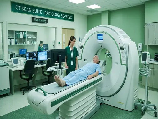
What Does a CT Scanner Look Like?
A CT scanner generally has a large circular opening.
Unlike many MRI scanners, the patient does not usually remain within a long tunnel.
The examination table moves through the opening while images are obtained.
Many patients therefore find CT easier to tolerate than MRI when significant claustrophobia is present.
Staying Still During CT
Movement can reduce image quality.
Patients may be asked to remain still for a short period.
For chest or abdominal scans, the technologist may give instructions such as:
-
“Breathe in.”
-
“Hold your breath.”
-
“Breathe normally.”
Following these instructions helps reduce motion on the images.
How Long Does a CT Scan Take?
CT imaging itself can often be performed relatively quickly.
However, the complete appointment may take longer because of:
-
Registration
-
Preparation
-
Oral contrast where required
-
IV placement
-
Kidney-function review
-
Contrast administration
-
Observation after contrast where appropriate
The exact duration depends on the examination.
Patients should ask about their specific CT rather than relying on one universal scan time.
After CT Without Contrast
After a routine CT without contrast, most patients can usually return to normal activity immediately unless their medical condition requires other restrictions.
The Radiology team will provide any specific instructions.
After Contrast-Enhanced CT
After IV contrast, patients may receive additional instructions according to:
-
Medical condition
-
Kidney function
-
Contrast reaction history
-
Other clinical factors
Patients should seek medical advice if they develop significant symptoms after leaving the imaging department.
CT Results
CT images are reviewed by a radiologist or another appropriately qualified imaging specialist.
The report may describe:
-
Normal structures
-
Abnormalities
-
Location
-
Size
-
Extent
-
Relationship to surrounding tissues
-
Comparison with previous imaging
-
Suggested further investigation
The final report should be interpreted in combination with the patient’s symptoms and medical history.
CT Images vs CT Report
A CT study may produce hundreds or thousands of images.
The radiologist reviews these images and creates a written report.
Patients may receive:
-
Electronic images
-
Printed or digital report
-
Other access to imaging records
depending on hospital systems.
Treatment decisions should generally be based on the radiologist’s interpretation together with the responsible clinician’s assessment.
Incidental Findings
CT may occasionally reveal an abnormality unrelated to the reason for scanning.
This is called an incidental finding.
Examples may include:
-
Cysts
-
Small nodules
-
Benign anatomical variations
-
Other unexpected abnormalities
Depending on the finding, the doctor may recommend:
-
No further action
-
Follow-up imaging
-
Another investigation
-
Specialist consultation
Not every incidental finding represents serious disease.
Abnormal CT Does Not Automatically Mean Surgery
CT can show abnormalities that may be managed:
-
Conservatively
-
With medication
-
Through monitoring
-
With another diagnostic test
-
Through surgery in selected cases
For example, detecting:
-
A disc abnormality
-
A kidney stone
-
A lung nodule
-
A gallbladder-related finding
does not automatically determine one specific treatment.
Clinical assessment remains essential.
Comparing Previous CT Scans
Previous imaging can help determine whether an abnormality is:
-
New
-
Stable
-
Increasing
-
Decreasing
-
Responding to treatment
Patients should provide previous CT or MRI examinations where available.
This can sometimes prevent unnecessary repeat imaging.
CT After Surgery
CT may be useful after selected operations when there is concern about:
-
Bleeding
-
Infection
-
Abscess
-
Fluid collection
-
Bowel complications
-
Tumor recurrence
-
Other postoperative problems
Routine CT is not required after every operation.
The surgeon determines when postoperative imaging is appropriate.
CT & Radiology
CT is part of the wider Radiology Service at AS Medical Complex.
The radiologist helps determine:
-
Appropriate CT protocol
-
Whether contrast is required
-
Scan area
-
Radiation optimization
-
Need for additional imaging
-
Interpretation
Providing an accurate clinical history helps the radiologist tailor the examination to the medical question.
CT & Emergency Department
CT plays a major role in Emergency care.
Depending on the presentation, emergency CT may be used for:
-
Head injury
-
Stroke assessment
-
Major trauma
-
Severe abdominal pain
-
Internal bleeding
-
Suspected pulmonary embolism
-
Other urgent conditions
CT should be integrated with:
-
Examination
-
Laboratory tests
-
Monitoring
-
Specialist consultation
rather than used independently.
CT & Neurology
Neurology may request CT in selected patients with:
-
Acute neurological symptoms
-
Stroke-related assessment
-
Headache with concerning features
-
Other neurological conditions
MRI may sometimes be recommended afterward when additional soft-tissue detail is required.
CT & Neurosurgery
Neurosurgery may use CT to evaluate:
-
Head trauma
-
Skull fractures
-
Intracranial bleeding
-
Hydrocephalus
-
Spine fractures
-
Postoperative conditions
-
Other neurosurgical problems
CT can be particularly useful for emergency and bony assessment.
CT & Pulmonology
Pulmonology may use CT or high-resolution CT for:
-
Lung nodules
-
Interstitial lung disease
-
Pulmonary fibrosis
-
Bronchiectasis
-
Complicated infection
-
Other lung conditions
CT findings are interpreted alongside:
-
Symptoms
-
Pulmonary examination
-
Chest X-ray
-
Pulmonary Function Tests
-
Bronchoscopy where required
CT & Gastroenterology
Gastroenterology may use abdominal or pelvic CT for selected:
-
Inflammatory conditions
-
Liver abnormalities
-
Pancreatic disease
-
Bowel disease
-
Complications
-
Other gastrointestinal conditions
Depending on the problem, patients may instead or additionally require:
-
Ultrasound
-
MRI
-
Endoscopy
-
Colonoscopy
-
Laboratory testing
CT & General Surgery
General & Laparoscopic Surgery may use CT when evaluating:
-
Appendicitis
-
Bowel obstruction
-
Abscess
-
Perforation
-
Trauma
-
Abdominal masses
-
Other surgical conditions
Emergency surgical decisions should be based on the complete clinical assessment.
CT & Urology
Urology may request CT for:
-
Kidney stones
-
Urinary obstruction
-
Kidney masses
-
Urinary trauma
-
Other selected conditions
The required CT protocol depends on the diagnostic question.
CT & Vascular Surgery
Vascular Surgery may use CT angiography in selected patients with:
-
Peripheral arterial disease
-
Aortic aneurysm
-
Arterial blockage
-
Selected vascular trauma
-
Other arterial conditions
Availability of advanced CTA protocols should be confirmed according to AS Medical Complex facilities.
CT & Orthopedics
Orthopedics may request CT for:
-
Complex fractures
-
Joint fractures
-
Bone abnormalities
-
Surgical planning
-
Selected postoperative assessment
MRI may be more appropriate when the main concern involves:
-
Ligaments
-
Tendons
-
Muscles
-
Other soft tissues
CT & Oncology
CT plays an important role in many cancer pathways.
It may be used for:
-
Tumor assessment
-
Staging
-
Treatment planning
-
Monitoring treatment
-
Follow-up
Depending on the cancer, additional assessment may involve:
-
MRI
-
Ultrasound
-
Endoscopy
-
Biopsy
-
Pathology
-
Other imaging
CT Limitations
CT is highly useful, but it has limitations.
These include:
-
Exposure to ionizing radiation
-
Contrast-related risks when contrast is used
-
Less soft-tissue contrast than MRI for some conditions
-
Incidental findings
-
Need for additional testing in selected cases
CT should therefore be selected because it is medically appropriate rather than simply because it is available.
When Should You Consider a CT Scan?
You may need CT assessment if your doctor is investigating:
-
Significant head injury
-
Acute neurological symptoms
-
Chest disease
-
Lung abnormalities
-
Severe abdominal symptoms
-
Kidney stones
-
Major trauma
-
Complex fracture
-
Suspected internal bleeding
-
Selected vascular disease
-
Tumor
-
Infection
-
Abnormal findings on another scan
-
Postoperative complications
-
Other clinically appropriate conditions
The CT examination should ideally answer a specific diagnostic question.
When Should Contrast Safety Be Reviewed?
Tell the Radiology team before a contrast-enhanced CT if you have:
-
Previous iodinated contrast reaction
-
Significant allergies
-
Chronic Kidney Disease
-
Kidney failure
-
Recent acute kidney injury
-
Dialysis
-
Diabetes
-
Pregnancy
-
Significant current illness
-
Other relevant medical conditions
The Radiology team can determine whether:
-
Contrast can be given normally
-
Additional testing is required
-
Precautions are required
-
Another imaging method should be considered
When Is Emergency Care Needed Instead of Routine CT?
Do not wait for a routine CT appointment if you develop:
-
Sudden facial weakness
-
Sudden arm or leg weakness
-
Sudden difficulty speaking
-
Severe chest pain
-
Sudden severe shortness of breath
-
Major trauma
-
Loss of consciousness
-
Severe uncontrolled bleeding
-
Sudden severe abdominal pain with collapse
-
Severe neurological deterioration
-
Other potentially life-threatening symptoms
These situations require urgent clinical assessment.
The Emergency team will determine whether CT, MRI, ultrasound, X-ray, or another investigation is required.
Multidisciplinary CT Care
Depending on CT findings, patient care may be coordinated with:
-
Radiology
-
Emergency Medicine
-
General Medicine
-
Neurology
-
Neurosurgery
-
Pulmonology
-
Gastroenterology
-
General & Laparoscopic Surgery
-
Orthopedics
-
Ortho & Spine Surgery
-
Urology
-
Vascular Surgery
-
Cardiology
-
Gynecology
-
Oncology
-
Nephrology
-
ENT
-
Other relevant specialties
CT is therefore often one stage within a broader diagnostic and treatment pathway.
Follow-Up After CT
The next step after CT depends on the findings.
Possible outcomes include:
-
No further investigation
-
Medical treatment
-
Specialist referral
-
Follow-up CT
-
MRI
-
Ultrasound
-
Endoscopy
-
Colonoscopy
-
Biopsy
-
Surgery
-
Other investigations
Significant findings should be reviewed with the doctor responsible for the patient’s care.
Repeat CT
Repeat CT may sometimes be required when:
-
A known disease needs monitoring
-
Symptoms change
-
Treatment response needs evaluation
-
Cancer surveillance is required
-
Postoperative complications are suspected
-
Another clinical indication develops
Because CT uses ionizing radiation, repeat examinations should have a valid medical indication.
Previous scans should be reviewed where possible before another CT is ordered.
CT Scan FAQs
What is a CT Scan?
A CT Scan – Computed Tomography – is a diagnostic imaging examination that uses specialized X-rays and computer processing to create detailed cross-sectional images of structures inside the body. CT can show bones, organs, blood vessels, lungs, and soft tissues in considerably greater detail than many routine X-rays.
Does a CT Scan use radiation?
Yes. CT uses ionizing X-ray radiation. The amount depends on the examination and scan protocol. Although radiation exposure carries a small potential long-term risk, medically necessary CT can provide important diagnostic information, and the expected benefit generally outweighs the small radiation risk when the examination is appropriately justified.
Does every CT Scan require contrast?
No. Many CT examinations can be performed without intravenous contrast. Contrast may be used when it improves visualization of blood vessels, organs, tumors, inflammation, infection, or other abnormalities. The radiologist determines whether contrast is necessary for the specific clinical question.
Can I have contrast CT if I have kidney disease?
Possibly. Kidney disease does not automatically prevent contrast-enhanced CT, but the Radiology team may need to review kidney function, recent creatinine or eGFR, dialysis status, clinical urgency, and alternative imaging before iodinated contrast is administered. Patients with known kidney disease should always inform the imaging team beforehand.
Can a pregnant woman have a CT Scan?
CT uses ionizing radiation, so pregnancy should always be disclosed and the examination should be clinically justified. However, necessary CT should not automatically be withheld when important or urgent diagnostic information is required. The doctor and radiologist should consider the body area, expected radiation exposure, urgency, pregnancy stage, and possible alternative imaging.
How long does a CT Scan take?
The actual CT image acquisition is often relatively quick, but the complete appointment may take longer if preparation, oral contrast, IV placement, kidney-function review, or contrast administration is required. The total time therefore depends on the exact examination.
Which is better: CT or MRI?
Neither is universally better. CT is often preferred for rapid emergency imaging, trauma, acute bleeding, lung disease, kidney stones, and detailed bone assessment. MRI can provide better soft-tissue contrast for many brain, spinal cord, nerve, ligament, tendon, muscle, and joint conditions and does not use ionizing radiation. The correct test depends on the clinical question.
CT Scan Services at AS Medical Complex
The CT Scan Service at AS Medical Complex is designed to provide rapid and detailed diagnostic imaging for appropriately selected patients.
From CT of the brain, head, chest, lungs, abdomen, pelvis, spine, bones, urinary system, and trauma assessment to contrast-enhanced CT, selected vascular imaging, cancer assessment, infection evaluation, postoperative imaging, and emergency diagnostic support, our goal is to provide clinically useful imaging that assists physicians in making informed treatment decisions.
Patient safety remains central to CT imaging. Every examination should balance the diagnostic benefit against radiation exposure, while patients undergoing contrast-enhanced CT should receive appropriate assessment of previous contrast reactions, kidney disease, pregnancy, diabetes, and other relevant medical conditions.
CT is particularly valuable because of its speed and ability to evaluate many internal structures simultaneously, but it should not automatically replace MRI, ultrasound, X-ray, or other investigations when those methods are more appropriate.
CT findings can be coordinated with the relevant departments at AS Medical Complex, including Emergency Services, Neurology, Neurosurgery, Pulmonology, Gastroenterology, General & Laparoscopic Surgery, Orthopedics, Urology, Vascular Surgery, Oncology, Nephrology, and other specialties where clinically appropriate.
Book a CT Scan at AS Medical Complex for physician-requested brain, head, chest, lung, abdominal, pelvic, spinal, bone, trauma, urinary, vascular, contrast-enhanced, and other clinically indicated CT examinations.